. 2015 Nov;75(15):1704-17.
doi: 10.1002/pros.23051. Epub 2015 Aug 7.

Relationship between serum response factor and androgen receptor in prostate cancer

Affiliations

Relationship between serum response factor and androgen receptor in prostate cancer

Maria Prencipe et al. Prostate. 2015 Nov.

Abstract

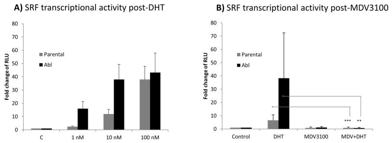
Background: Serum response factor (SRF) is an important transcription factor in castrate-resistant prostate cancer (CRPC). Since CRPC is associated with androgen receptor (AR) hypersensitivity, we investigated the relationship between SRF and AR.

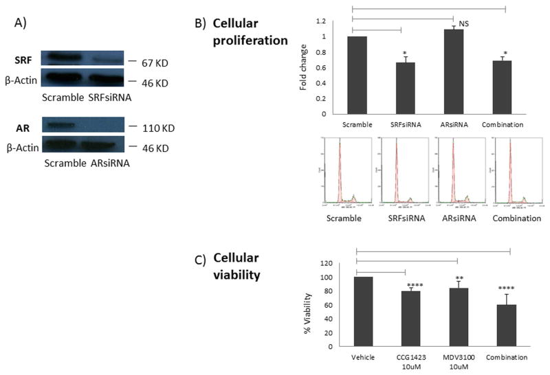
Materials and methods: Transcriptional activity was assessed by luciferase assay. Cell proliferation was measured by MTT and flow cytometry. Protein expression in patients was assessed by immunohistochemistry.

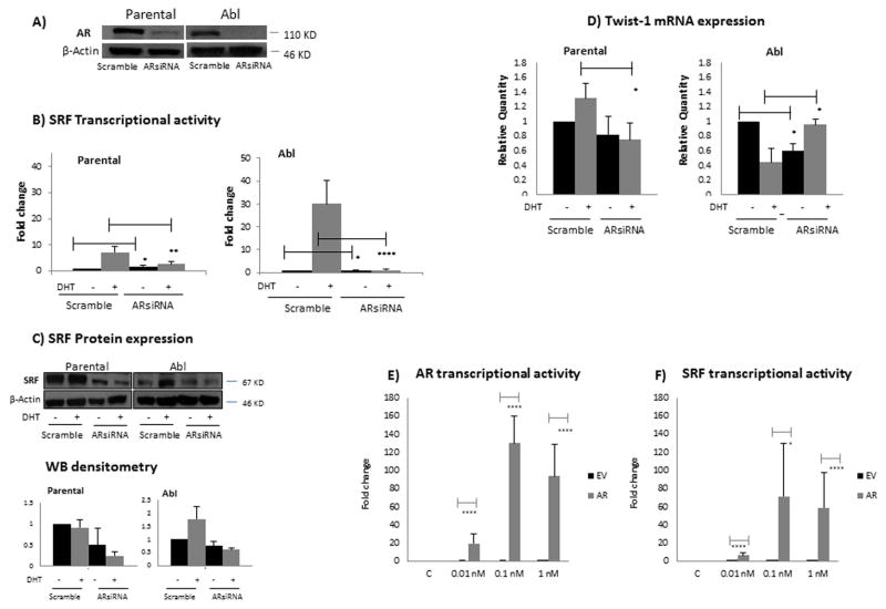
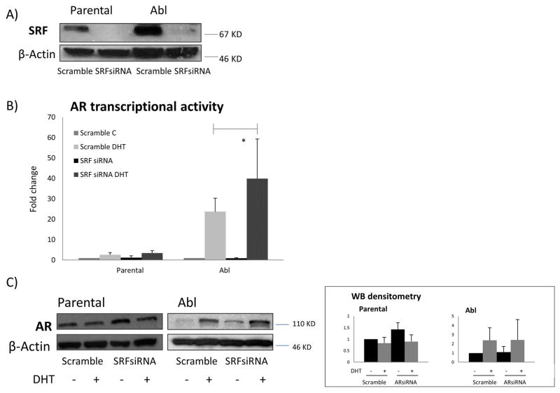
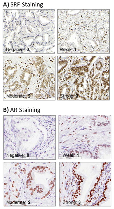
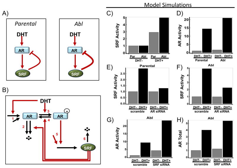
Results: To investigate AR involvement in SRF response to androgen, AR expression was down-regulated using siRNA. This resulted in the abrogation of SRF induction post-DHT. Moreover, DHT stimulation failed to induce SRF transcriptional activity in AR-negative PC346 DCC cells, which was only restored following AR over-expression. Next, SRF expression was down-regulated by siRNA, resulting in AR increased transcriptional activity in castrate-resistant LNCaP Abl cells but not in the parental LNCaP. This negative feedback loop in the resistant cells was confirmed by immunohistochemistry which showed a negative correlation between AR and SRF expression in CRPC bone metastases and a positive correlation in androgen-naïve prostatectomies. Cell proliferation was next assessed following SRF inhibition, demonstrating that SRF inhibition is more effective than AR inhibition in castrate-resistant cells.

Conclusion: Our data support SRF as a promising therapeutic target in combination with current treatments.

Keywords: androgen receptor; castrate-resistant prostate cancer; serum response factor.


Figures

Figure 1
Figure 1. SRF transcriptional activity in response to androgen stimulation and inhibition
Cells were seeded in 12 well plates at a density of 5×10⁵ cells per well. The following day they were transiently transfected with the reporter construct pGL4.34 driven by a SRF responsive element (CArG box) that drives the transcription of the luciferase reporter gene luc2P. Twenty four hours post-transfection cells were treated with increasing concentrations of DHT (1, 10 and 100 nM) (panel A) or 10 μM of MDV3100 alone, 10 nM of DHT alone, MDV3100 and DHT in combination (panel B). Reporter gene activity was measured 24hours after treatment. Columns, mean values obtained from three independent experiments in triplicate; bars, SD. Mean values were compared using t-test assuming equal variances. ** p<0.01; *** p<0.001. RLU: relative luciferase units.
Figure 2
Figure 2. SRF transcriptional activity is dependent on AR
A) Cells were seeded in 6 well plates at a density of 250,000 cells per well. The following day they were transiently transfected with a pool of siRNAs specific for AR or non- specific control siRNAs. Following 48 hours, total protein extracts were prepared for western blotting and β-actin was used as loading control. Representative images from three independent experiments are shown. B) Cells were seeded in 12 well plates at a density of 5×10⁵ cells per well. The following day they were transiently transfected with a pool of siRNAs specific for AR or non- specific control siRNAs. Twenty four hours post-siRNA transfection, cells were transfected again with the reporter construct driven by an SRF responsive element. The following day cells were either left untreated or treated with 10 nM DHT. Reporter gene activity was measured 24 hours after treatment. Columns, mean values obtained from three independent experiments in triplicate; bars, SD. Mean values were compared using t-test assuming equal variances. * p<0.05, ** p<0.01, **** p<1 E-04. C) Cells were seeded in 6 well plates at a density of 250,000 cells per well. The following day they were transiently transfected with a pool of siRNAs specific for AR or non- specific control siRNAs. Twenty four hours post-siRNA transfection, cells were either left untreated or treated with 10 nM DHT. Total protein extracts were prepared for western blotting 24 hours after treatment. β-actin was used as loading control. Representative images from three independent experiments are shown. Densitometry is shown on the right panel. Columns, mean values obtained from three independent experiments in triplicate; bars, SD. D) Cells were seeded and treated as in panel C. Forty-eight hours after DHT treatment, RNA was isolated and cDNA was synthesised. The cDNA was subsequently used as template for gene-specific real time quantitative reverse transcription PCR (qRT-PCR). Columns, mean values obtained from three independent experiments in triplicate; bars, SD. Mean values were compared using t-test assuming equal variances. * p<0.05 E-F) PC346 DCC cells were seeded in 12 well plates at a density of 5×10⁵ cells per well. The following day they were transiently transfected either with a pEGFP-C1-AR plasmid or with the empty vector (EV). Twenty four hours post-siRNA transfection, cells were transfected again with either the reporter construct driven by an AR responsive element (panel D) or with the reporter construct driven by a SRF responsive element (panel E). The following day cells were treated with increasing concentrations (0.01, 0.1, 1 nM) of DHT. Reporter gene activity was measured 24 hours after treatment. Black columns, EV; grey columns, pEGFP-C1-AR plasmid. Columns, mean values obtained from three independent experiments in triplicate; bars, SD. Mean values were compared using t-test assuming equal variances. * p<0.05, **** p<1 E-04.
Figure 3
Figure 3. AR transcriptional activity is affected by SRF
A) Cells were seeded in 6 well plates at a density of 250,000 cells per well. The following day they were transiently transfected with a pool of siRNAs specific for SRF or non- specific control siRNAs. Following 48 hours, total protein extracts were prepared for western blotting and β-actin was used as loading control. Representative images from three independent experiments are shown. B) Cells were seeded in 12 well plates at a density of 5×10⁵ cells per well. The following day they were transiently transfected with a pool of siRNAs specific for SRF or non- specific control siRNAs. Twenty four hours post-siRNA transfection, cells were transfected again with the reporter construct driven by an AR responsive element. The following day cells were either left untreated or treated with 10 nM DHT. Reporter gene activity was measured 24 hours after treatment. Columns, mean values obtained from three independent experiments in triplicate; bars, SD. Mean values were compared using t-test assuming equal variances. * p<0.05. C) Cells were seeded in 6 well plates at a density of 250,000 cells per well. The following day they were transiently transfected with a pool of siRNAs specific for SRF or non- specific control siRNAs. Twenty four hours post-siRNA transfection, cells were either left untreated or treated with 10 nM DHT. Total protein extracts were prepared for western blotting 24 hours after treatment. β-actin was used as loading control. Representative images from three independent experiments are shown. Densitometry is shown on the right panel. Columns, mean values obtained from three independent experiments in triplicate; bars, SD.
Figure 4
Figure 4. Protein expression assessed by IHC on clinical tissues from patients with prostate cancer
A) SRF IHC staining. B) AR IHC staining. Examples of negative (1), weak (2), moderate (3) and strong (4) nuclear staining are shown for both SRF and AR (40X magnifications).
Figure 5
Figure 5. Impact of SRF inhibition on cellular viability and proliferation
A) Cells were seeded in 6 well plates at a density of 250,000 cells per well. The following day they were transiently transfected with a pool of siRNAs specific for AR, SRF or non- specific control siRNAs. Following 48 hours, total protein extracts were prepared for western blotting and β-actin was used as loading control. Representative images from three independent experiments are shown. B) Cells were seeded in 6 well plates at a density of 250,000 cells per well. The following day they were transiently transfected either with a non- specific control siRNAs, a pool of siRNAs specific for SRF, a pool of siRNAs specific for AR or a combination of SRF-siRNA and AR- siRNA. Following 48 hours, cells were tripsinised and stained with PI prior to FACS analysis. Proliferation was assessed by measuring the S phase within the cell cycle. C) Cells were seeded in 12 well plates at a density of 5×10⁵ cells per well. The following day they were treated either with vehicle (DMSO), 10 μM of CCG-1423, 10 μM of MDV3100 or a combination of CCG-1423 and MDV3100. Following 48 hours, cellular viability was measured by MTT assay. Columns, mean values obtained from three independent experiments in triplicate; bars, SD. Mean values were compared using t-test assuming equal variances. * p<0.05; ** p<0.01; **** p<1 E-04.
Figure 6
Figure 6. Computational model and simulations of the proposed mechanism of SRF action
A) A simplified diagram showing the existence of a negative feedback mechanism between SRF and AR under DHT stimulation. B) An assumed kinetic scheme describing the mechanistic reactions of the AR-SRF network that capture the negative feedback regulation (see Supplementary Information for details). C) Model simulations of SRF activity comparing scramble and AR knock-down using siRNA. D–E) Model simulations of AR activity and expression level comparing scramble and SRF knock-down using siRNA.

References

    1. Prencipe M, Madden SF, O’Neill A, O’Hurley Gillian, Culhane Aedin, O’Connor Darran, Klocker H, Kay EW, Gallagher WM, Watson WR. Identification of transcription factors associated with castration-resistance: is the Serum Responsive Factor a potential therapeutic target? Prostate. 2013;73(7):743–53. - PubMed
    1. Sun Q, Chen G, Streb JW, Long X, Yang Y, Stoeckert CJ, Jr, Miano JM. Defining the mammalian CArGome. Genome research. 2006;16(2):197–207. - PMC - PubMed
    1. Vickers ER, Kasza A, Kurnaz IA, Seifert A, Zeef LA, O’donnell A, Hayes A, Sharrocks AD. Ternary complex factor-serum response factor complex-regulated gene activity is required for cellular proliferation and inhibition of apoptotic cell death. Molecular and cellular biology. 2004;24(23):10340–51. - PMC - PubMed
    1. Heemers HV, Regan KM, Dehm SM, Tindall DJ. Androgen induction of the androgen receptor coactivator four and a half LIM domain protein-2: evidence for a role for serum response factor in prostate cancer. Cancer Res. 2007;67(21):10592–9. - PubMed
    1. O’Hurley G, Prencipe M, Lundon D, O’Neill A, Boyce S, O’Grady A, Gallagher WM, Morrissey C, Kay EW, Watson W. The analysis of Serum Response Factor expression in bone and soft tissue Prostate Cancer metastases. Prostate. 2014;74:306–313. - PubMed

Publication types

MeSH terms