. 2015 Aug 7:5:12895.
doi: 10.1038/srep12895.

Sex hormones have pervasive effects on thymic epithelial cells

Affiliations

Sex hormones have pervasive effects on thymic epithelial cells

Maude Dumont-Lagacé et al. Sci Rep. .

Abstract

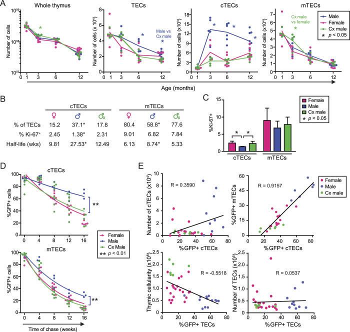
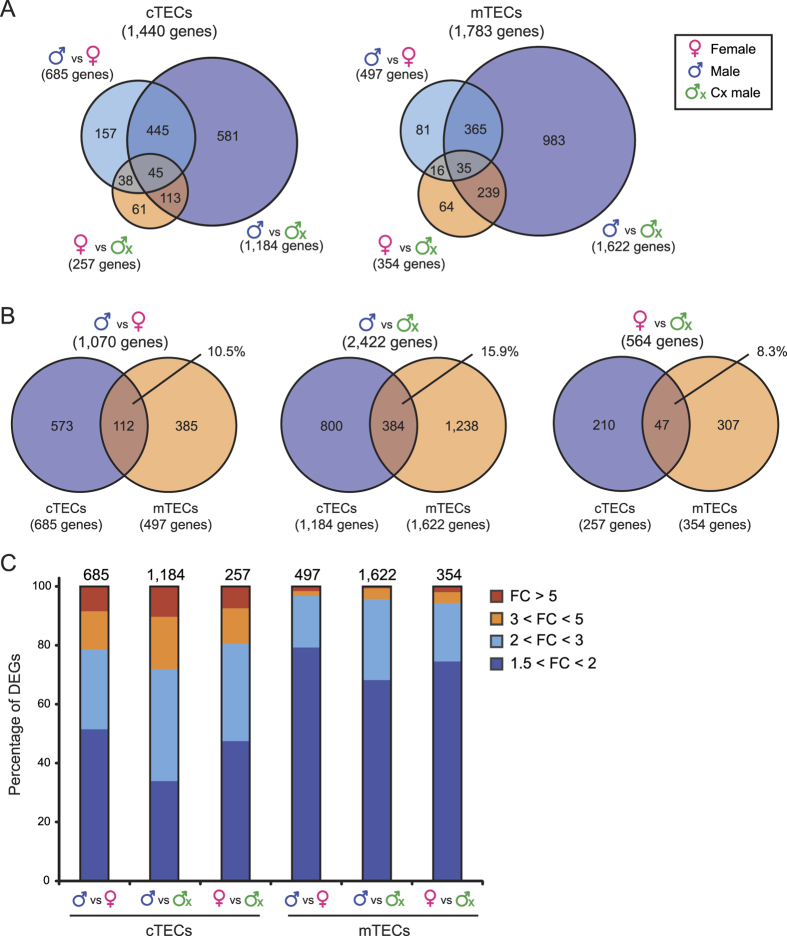
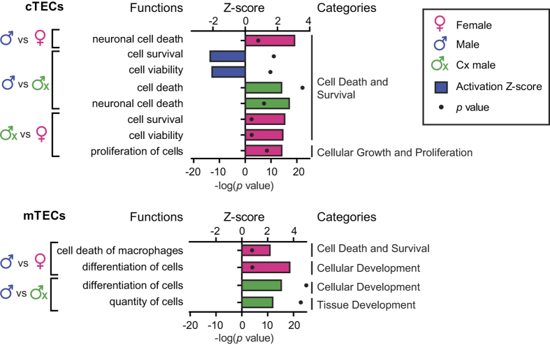
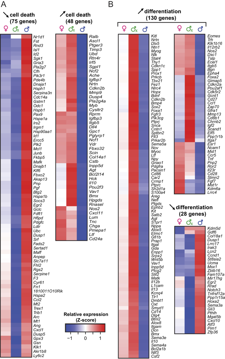
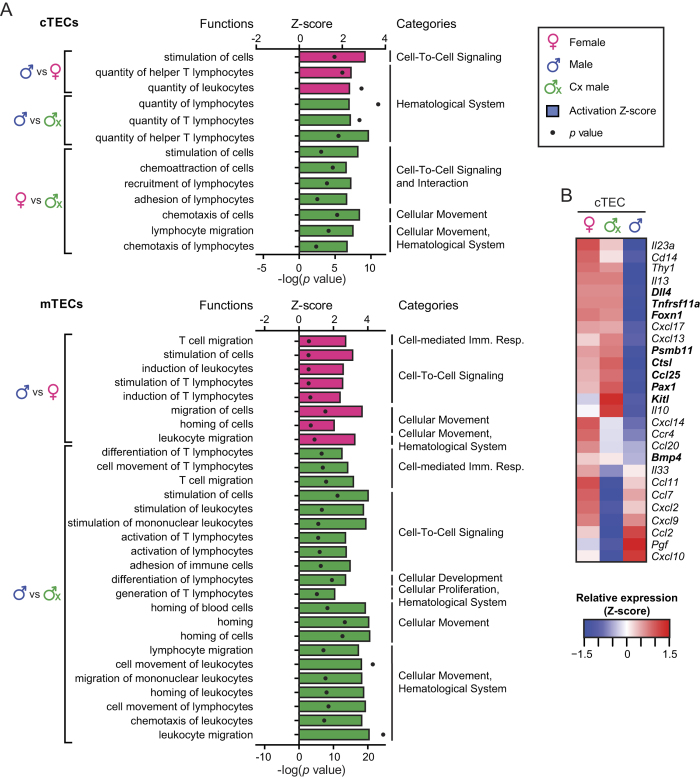
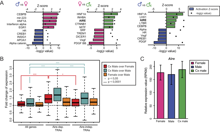
The goal of our study was to evaluate at the systems-level, the effect of sex hormones on thymic epithelial cells (TECs). To this end, we sequenced the transcriptome of cortical and medullary TECs (cTECs and mTECs) from three groups of 6 month-old mice: males, females and males castrated at four weeks of age. In parallel, we analyzed variations in the size of TEC subsets in those three groups between 1 and 12 months of age. We report that sex hormones have pervasive effects on the transcriptome of TECs. These effects were exquisitely TEC-subset specific. Sexual dimorphism was particularly conspicuous in cTECs. Male cTECs displayed low proliferation rates that correlated with low expression of Foxn1 and its main targets. Furthermore, male cTECs expressed relatively low levels of genes instrumental in thymocyte expansion (e.g., Dll4) and positive selection (Psmb11 and Ctsl). Nevertheless, cTECs were more abundant in males than females. Accumulation of cTECs in males correlated with differential expression of genes regulating cell survival in cTECs and cell differentiation in mTECs. The sexual dimorphism of TECs highlighted here may be mechanistically linked to the well-recognized sex differences in susceptibility to infections and autoimmune diseases.


Figures

Figure 1
Figure 1. Male mice show an accumulation of cTECs with age that is not due to enhanced cell proliferation.
(a) Thymic cell populations in mice aged from 1 to 12 months. The gating strategy is depicted in Supplementary Figure S1c. (b) Percentage of TEC populations in 6 month-old mice, mean percentage of Ki-67+ cells at 4–5 months of age, and half-life (in weeks) of H2B-GFP+ TECs calculated using one phase decay nonlinear regression. (c) Percentages of Ki-67+ TECs in 4 to 5 month-old mice. (d) H2B-GFP dilution in adult mice during a chase period of 16 weeks. (e) Correlation (Pearson) between different features of TEC populations in 6 month-old mice. Each dot represents onemouse.
Figure 2
Figure 2. Transcriptomic signatures of cTECs and mTECs in 6 month-old male, female and Cx male mice.
(a) Venn diagram representation of DEGs in cTECs and mTECs from the three experimental groups. (b) Overlap between DEGs in cTECs and mTECs. (c) Percentages of DEGs for selected thresholds of mRNA expression fold-change (FC). Numbers of DEGs are indicated above bars.
Figure 3
Figure 3. IPA analysis of DEGs in cTECs and mTECs.
IPA analysis of DEGs predicts decreased cell death and cell differentiation in males. Activation Z-scores are depicted with bars, whereas p values are shown with black dots. The color of bars shows in which group a given process is activated (e.g., blue = males). All functions represented on the graph were significantly enriched (p < 0.05).
Figure 4
Figure 4. Relative expression of genes contributing to the inhibition of cell death and cell differentiation in male TECs.
(a) Relative expression of genes that affect cell death in cTECs. (b) Relative expression of genes that affect cell differentiation in mTECs. Relative gene expression is depicted as a Z-score, calculated separately for cTECs and mTECs. Red corresponds to higher expression, whereas blue corresponds to lower expression.
Figure 5
Figure 5. Several TEC genes involved in thymopoiesis are downregulated in male TECs.
(a) IPA analysis of DEGs predicts differential activation of biological functions related to homing, stimulation of cells, chemotaxis and production of T lymphocytes. Activation Z-score is depicted with bars, whereas p values are shown with black dots. The color of bars shows in which group a given process is activated (e.g., green = Cx males). All functions represented on the graph are significantly enriched (p < 0.05). (b) Heatmaps of relative cTEC expression of IPA-discovered genes that may affect thymopoiesis. Genes that have well-characterized functions in the thymus are highlighted in bold. The gene expression is depicted as a Z-score, calculated separately for each cell type. Red corresponds to higher expression, whereas blue corresponds to lower expression.
Figure 6
Figure 6. Cx males show higher expression of TRAs.
(a) Ten most activated upstream regulators predicted by IPA analysis of mTECs DEGs in female vs male (left), in Cx male vs female (center) and in Cx male vs male (right, all predicted activators shown are significant p < 0.05). The color of bars shows in which group a given upstream regulator is activated (e.g., blue = males). (b) Fold-difference in expression of all genes, Aire-dependent TRAs and Aire-independent TRAs in mTECs. The gene expression ratio (log2 RPKM) of Cx male over female is depicted in red, of Cx male over male in turquoise and of female over male in orange. (c) Relative expression of Aire in mTECs (RPKM).

References

    1. Whitacre C. C. Sex differences in autoimmune disease. Nat Immunol 2, 777–780 (2001). - PubMed
    1. Fish E. N. The X-files in immunity: sex-based differences predispose immune responses. Nat Rev Immunol 8, 737–744 (2008). - PMC - PubMed
    1. Markle J. G. & Fish E. N. SeXX matters in immunity. Trends Immunol. 35, 97–104 (2014). - PubMed
    1. Danska J. S. Sex matters for mechanism. Sci Transl Med 6, 258fs40 (2014). - PubMed
    1. Markle J. G. et al. Sex differences in the gut microbiome drive hormone-dependent regulation of autoimmunity. Science 339, 1084–1088 (2013). - PubMed

Publication types

Substances