. 2015 Oct;36(10):1866-73.
doi: 10.3174/ajnr.A4387. Epub 2015 Aug 6.

Differentiation between Cystic Pituitary Adenomas and Rathke Cleft Cysts: A Diagnostic Model Using MRI

Affiliations

Differentiation between Cystic Pituitary Adenomas and Rathke Cleft Cysts: A Diagnostic Model Using MRI

M Park et al. AJNR Am J Neuroradiol. 2015 Oct.

Abstract

Background and purpose: Cystic pituitary adenomas may mimic Rathke cleft cysts when there is no solid enhancing component found on MR imaging, and preoperative differentiation may enable a more appropriate selection of treatment strategies. We investigated the diagnostic potential of MR imaging features to differentiate cystic pituitary adenomas from Rathke cleft cysts and to develop a diagnostic model.

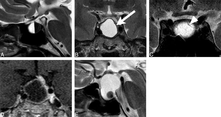
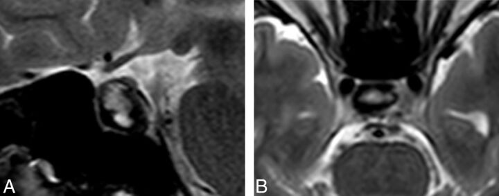
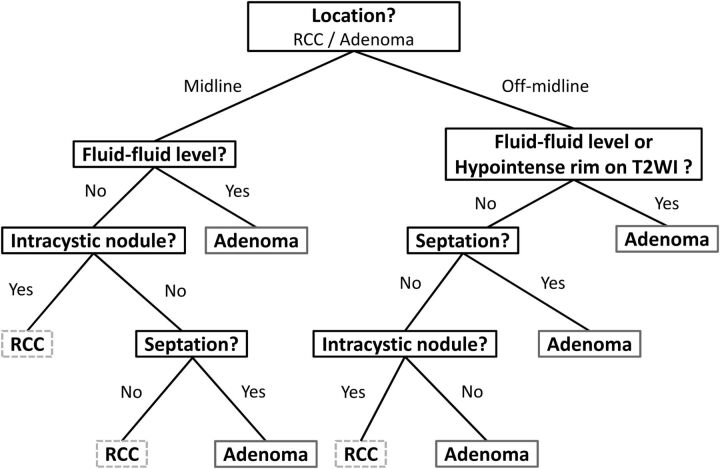
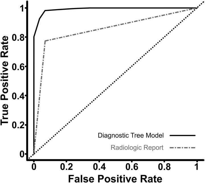
Materials and methods: This retrospective study included 54 patients with a cystic pituitary adenoma (40 women; mean age, 37.7 years) and 28 with a Rathke cleft cyst (18 women; mean age, 31.5 years) who underwent MR imaging followed by surgery. The following imaging features were assessed: the presence or absence of a fluid-fluid level, a hypointense rim on T2-weighted images, septation, an off-midline location, the presence or absence of an intracystic nodule, size change, and signal change. On the basis of the results of logistic regression analysis, a diagnostic tree model was developed to differentiate between cystic pituitary adenomas and Rathke cleft cysts. External validation was performed for an additional 16 patients with a cystic pituitary adenoma and 8 patients with a Rathke cleft cyst.

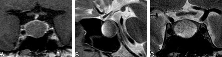
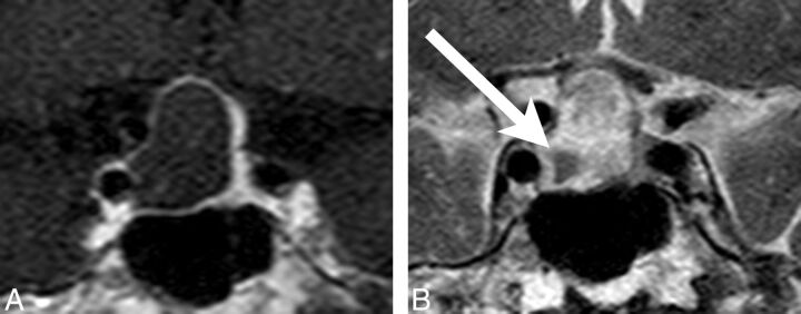
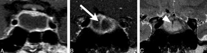
Results: The presence of a fluid-fluid level, a hypointense rim on T2-weighted images, septation, and an off-midline location were more common with pituitary adenomas, whereas the presence of an intracystic nodule was more common with Rathke cleft cysts. Multiple logistic regression analysis showed that cystic pituitary adenomas and Rathke cleft cysts can be distinguished on the basis of the presence of a fluid-fluid level, septation, an off-midline location, and the presence of an intracystic nodule (P = .006, .032, .001, and .023, respectively). Among 24 patients in the external validation population, 22 were classified correctly on the basis of the diagnostic tree model used in this study.

Conclusions: A systematic approach using this diagnostic tree model can be helpful in distinguishing cystic pituitary adenomas from Rathke cleft cysts.


Figures

Fig 1.
Fig 1.
Typical MR features for image analysis: A, fluid-fluid level (sagittal T2-weighted image); B, peripheral hypointense rim (arrow) (coronal T2-weighted image); C, septation (arrowhead) (coronal T2-weighted image); D, off-midline location, deviating pituitary stalk (coronal contrast-enhanced T1-weighted image); and E, intracystic nodule (sagittal T2-weighted image).
Fig 2.
Fig 2.
Representative case of discordant interpretation. A, A hypointense rim on a T2-weighted image of a 27-year-old woman with a pituitary adenoma, which was misinterpreted as a fluid-fluid level on axial T2-weighted images by one reader. B, Later, a hypointense rim on a T2-weighted image was agreed on consensually.
Fig 3.
Fig 3.
Diagnostic decision tree for the differentiation of cystic pituitary adenomas and RCCs using MR imaging.
Fig 4.
Fig 4.
Receiver operating characteristic curve comparison between the diagnostic decision tree model and official radiologic report for the differentiation of cystic pituitary adenomas and Rathke cleft cysts.
Fig 5.
Fig 5.
A 71-year-old female patient presented with a 21.2-mm nonenhancing intrasellar cystic lesion. The lesion was located in the midline on a coronal contrast-enhanced T1-weighted image (A), and a fluid-fluid level (B) and septation (C) were seen on sagittal and coronal T2-weighted images. This lesion was classified as a cystic pituitary adenoma on the basis of the diagnostic tree model and was finally diagnosed by histopathology as a pituitary adenoma.
Fig 6.
Fig 6.
A 42-year-old male patient presented with a 23.8-mm nonenhancing sellar and suprasellar lesion. The cystic lesion had an off-midline location on contrast-enhanced T1-weighted imaging (A) and an intracystic nodule (arrow) showing hypointensity on coronal T2-weighted imaging (B). There was no identified fluid-fluid level or septation. Therefore, this lesion was classified as a Rathke cleft cyst on the basis of the diagnostic tree model and was diagnosed through histopathology as a Rathke cleft cyst.
Fig 7.
Fig 7.
A 19-year-old man presented with a nonenhancing intrasellar lesion with a midline location (A) and internal septation (B [arrow] and C [arrowhead]) on coronal contrast-enhanced T1-weighted imaging (A and B) and coronal T2-weighted imaging (C). There was no visible fluid-fluid level or intracystic nodule, so this lesion was classified as a cystic pituitary adenoma based of the diagnostic tree model; however, it was finally diagnosed as a Rathke cleft cyst.

Comment in

  • Hemorrhagic Pituitary Adenoma versus Rathke Cleft Cyst: A Frequent Dilemma.
    Bonneville JF. Bonneville JF. AJNR Am J Neuroradiol. 2016 Mar;37(3):E27-8. doi: 10.3174/ajnr.A4653. Epub 2015 Dec 17. AJNR Am J Neuroradiol. 2016. PMID: 26680456 Free PMC article. No abstract available.
  • Reply.
    Park M, Ahn SS. Park M, et al. AJNR Am J Neuroradiol. 2016 Mar;37(3):E29. doi: 10.3174/ajnr.A4668. Epub 2015 Dec 17. AJNR Am J Neuroradiol. 2016. PMID: 26680458 Free PMC article. No abstract available.

References

    1. Gutenberg A, Larsen J, Lupi I, et al. . A radiologic score to distinguish autoimmune hypophysitis from nonsecreting pituitary adenoma preoperatively. AJNR Am J Neuroradiol 2009;30:1766–72 10.3174/ajnr.A1714 - DOI - PMC - PubMed
    1. Rennert J, Doerfler A. Imaging of sellar and parasellar lesions. Clin Neurol Neurosurg 2007;109:111–24 10.1016/j.clineuro.2006.11.001 - DOI - PubMed
    1. Johnsen DE, Woodruff WW, Allen IS, et al. . MR imaging of the sellar and juxtasellar regions. Radiographics 1991;11:727–58 10.1148/radiographics.11.5.1947311 - DOI - PubMed
    1. Zhang X, Zhang W, Fu LA, et al. . Hemorrhagic pituitary macroadenoma: characteristics, endoscopic endonasal transsphenoidal surgery, and outcomes. Ann Surg Oncol 2011;18:246–52 10.1245/s10434-011-1809-x - DOI - PubMed
    1. Semple PL, Webb MK, de Villiers JC, et al. . Pituitary apoplexy. Neurosurgery 2005;56:65–72; discussion 72–73 - PubMed