

The Vulnerable Ventral Tegmental Area in Parkinson's Disease
- PMID: 26251824
- PMCID: PMC4523275
- DOI: 10.1016/j.baga.2015.06.001
The Vulnerable Ventral Tegmental Area in Parkinson's Disease
Abstract
Introduction: The involvement of dopaminergic neurons in the ventral tegmental area (VTA) in Parkinson's disease (PD) has not been universally recognized by neuroscientists and neurologists. Here, we conduct a review of previous research documenting dopaminergic neuronal loss in both the substantia nigra pars compacta (SNpc) and VTA and add three new post-mortem PD cases to the literature.
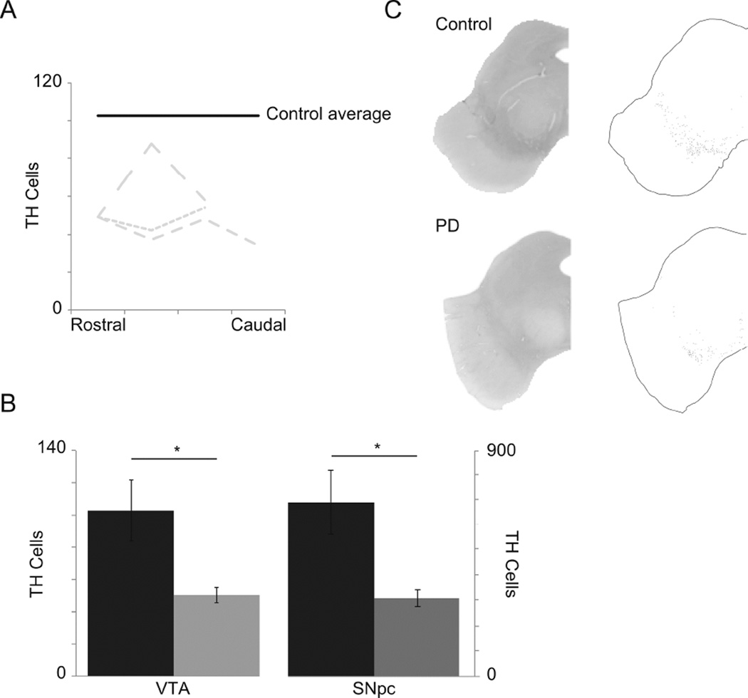
Methods: PD and control brains were sectioned, stained for tyrosine hydroxylase, and cells in the SNpc and VTA were counted.
Results: Based on the review, we report two main results: 1) the VTA does degenerate in PD, and 2) the VTA degenerates less than the SNpc.
Conclusion: Inconsistent clinical information about these cases limits our ability to interpret how the VTA contributes to PD symptoms. However, our data in combination with prior PD neuropathological cases in the literature unequivocally establish that the VTA is involved in PD, and could be relevant for future investigation of non-motor symptoms in PD.
Keywords: Parkinson’s disease; Ventral tegmental area.
Figures
References
-
- Davie CA. A review of Parkinson’s disease. Br Med Bull. 2008;86:109–127. - PubMed
-
- Jellinger KA. Pathology of Parkinson’s disease. Changes other than the nigrostriatal pathway. Mol Chem Neuropathol Spons Int Soc Neurochem World Fed Neurol Res Groups Neurochem Cerebrospinal Fluid. 1991;14:153–197. - PubMed
-
- Hur EE, Zaborszky L. Vglut2 afferents to the medial prefrontal and primary somatosensory cortices: a combined retrograde tracing in situ hybridization study [corrected] J Comp Neurol. 2005;483:351–373. - PubMed
-
- Lammel S, Hetzel A, Häckel O, Jones I, Liss B, Roeper J. Unique properties of mesoprefrontal neurons within a dual mesocorticolimbic dopamine system. Neuron. 2008;57:760–773. - PubMed
Grants and funding
LinkOut - more resources
Full Text Sources
Other Literature Sources
Miscellaneous