

Surgical Management in a Patient With Complex Uveitic Glaucoma: A Case Report
- PMID: 26252285
- PMCID: PMC4616600
- DOI: 10.1097/MD.0000000000001248
Surgical Management in a Patient With Complex Uveitic Glaucoma: A Case Report
Abstract
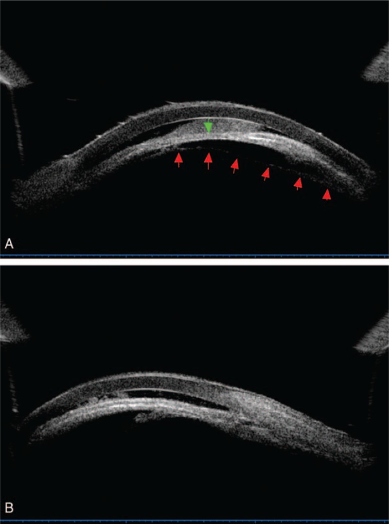
Uveitic glaucoma (UG) is secondary glaucoma, present as a clinical challenge in both diagnosis and management.We report a case of complex UG, which initially presented as pupillary block and rupture of the anterior lens capsule. We performed cataract extraction with preservation of posterior capsule. Then, the case turned to aphakic malignant glaucoma. We performed anterior vitrectomy with posterior capsule resection in this case. After the second operation, the patient had a satisfactory recovery. Specifically, ultrasonographic biomicroscopy was useful during the diagnosis process and follow-up period in this case.UG presenting as pupillary block, rupture of the anterior lens capsule, and aqueous misdirection seldom presents in clinical practice. Earlier and more active surgical intervention may be necessary for effective preservation of visual function in complex cases of UG.
Conflict of interest statement
The authors have no funding and conflicts of interest to disclose.
Figures


References
-
- Takahashi T, Ohtani S, Miyata K, et al. A clinical evaluation of uveitis-associated secondary glaucoma. Jpn J Ophthalmol 2002; 46:556–562. - PubMed
-
- Dietlein TS. Glaucoma uveitis. Causes of and treatment options for increased intraocular pressure in cases of inflammatory ophthalmology. Ophthalmologe 2003; 100:991–1006. - PubMed
-
- Siddique SS, Suelves AM, Baheti U, et al. Glaucoma and uveitis. Surv Ophthalmol 2013; 58:1–10. - PubMed
-
- Kishi A, Nao-i N, Sawada A. Ultrasound biomicroscopic findings of acute angle-closure glaucoma in Vogt-Koyanagi-Harada syndrome. Am J Ophthalmol 1996; 122:735–737. - PubMed
Publication types
MeSH terms
LinkOut - more resources
Full Text Sources
Medical
