. 2015 Aug 7;10(8):e0133937.
doi: 10.1371/journal.pone.0133937. eCollection 2015.

Peripheral Blood Mononuclear Cells Enhance Cartilage Repair in in vivo Osteochondral Defect Model

Affiliations

Peripheral Blood Mononuclear Cells Enhance Cartilage Repair in in vivo Osteochondral Defect Model

Niina Hopper et al. PLoS One. .

Abstract

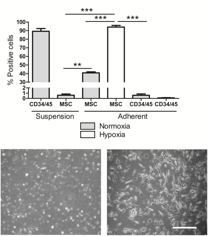
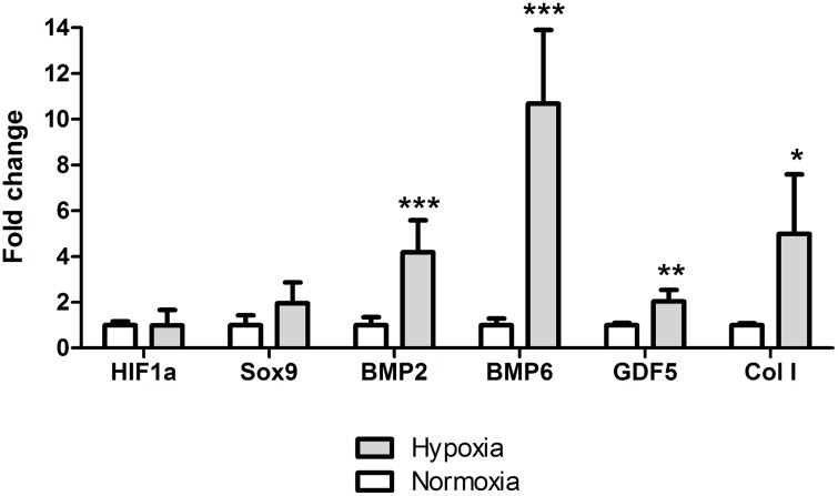
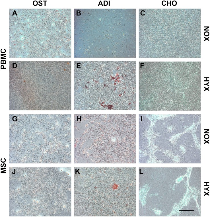
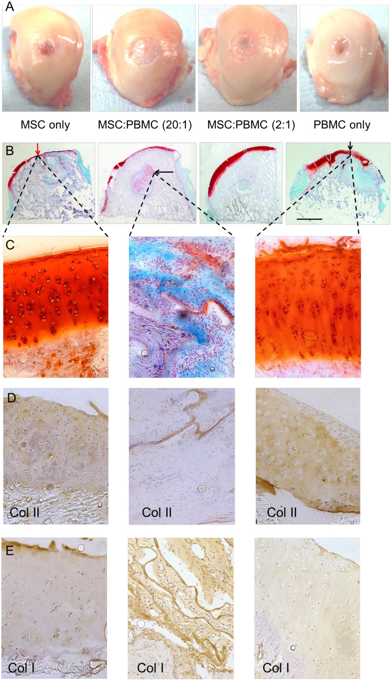
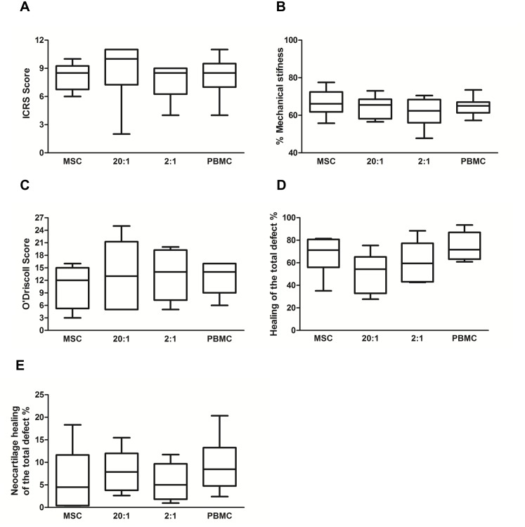
This study characterized peripheral blood mononuclear cells (PBMC) in terms of their potential in cartilage repair and investigated their ability to improve the healing in a pre-clinical large animal model. Human PBMCs were isolated with gradient centrifugation and adherent PBMC's were evaluated for their ability to differentiate into adipogenic, chondrogenic and osteogenic lineages and also for their expression of musculoskeletal genes. The phenotype of the PBMCs was evaluated using Stro-1, CD34, CD44, CD45, CD90, CD106, CD105, CD146 and CD166 cell surface markers. Osteochondral defects were created in the medial femoral condyle (MFC) of 24 Welsh mountain sheep and evaluated at a six month time point. Four cell treatment groups were evaluated in combination with collagen-GAG-scaffold: (1) MSC alone; (2) MSCs and PBMCs at a ratio of 20:1; (3) MSCs and PBMC at a ratio of 2:1 and (4) PBMCs alone. Samples from the surgical site were evaluated for mechanical properties, ICRS score and histological repair. Fresh PBMC samples were 90% positive for hematopoietic cell surface markers and negative for the MSC antibody panel (<1%, p = 0.006). However, the adherent PBMC population expressed mesenchymal stem cell markers in hypoxic culture and lacked CD34/45 positive cells (<0.2%). This finding demonstrated that the adherent cells had acquired an MSC-like phenotype and transformed in hypoxia from their original hematopoietic lineage. Four key genes in muskuloskeletal biology were significantly upregulated in adherent PBMCs by hypoxia: BMP2 4.2-fold (p = 0.0007), BMP6 10.7-fold (p = 0.0004), GDF5 2.0-fold (p = 0.002) and COL1 5.0-fold (p = 0.046). The monolayer multilineage analysis confirmed the trilineage mesenchymal potential of the adherent PBMCs. PBMC cell therapy was equally good as bone marrow MSC therapy for defects in the ovine large animal model. Our results show that PBMCs support cartilage healing and oxygen tension of the environment was found to have a key effect on the derivation of a novel adherent cell population with an MSC-like phenotype. This study presents a novel and easily attainable point-of-care cell therapy with PBMCs to treat osteochondral defects in the knee avoiding any cell manipulations outside the surgical room.


Conflict of interest statement

Competing Interests: The corresponding authors has read the journal's policy and the authors of this manuscript have the following competing interests: Prof. Neil Rushton has acted as paid consultant in the past five years for OrthoMimetics/Tigenix. None of the other authors have any conflict of interest to declare. This does not alter the authors’ adherence to PLOS ONE policies on sharing data and materials.

Figures

Fig 1
Fig 1. Peripheral blood mononuclear cell characterization.
(A) Fluorescent labelling of fresh PBMC in suspension and adherent PBMC in both normoxia and hypoxia comparing hematopoietic and mesenchymal cell surface markers (n = 4). Representative images of PBMCs after 12 days growing in (B) normoxia and (C) hypoxia (scale bar 50 μm).
Fig 2
Fig 2. Gene expression analysis.
The mRNA expression of PBMCs in both normoxic and hypoxic culture (24h). BMP2 (p = 0.0007), BMP6 (p = 0.0004), GDF5 (p = 0.002) and COL1 (p = 0.046) normalized to B2M housekeeping gene. Level of statistical significance; * p<0.05, ** p<0.001 and *** p<0.0001 with biological n = 4 and technical n = 3.
Fig 3
Fig 3. Tripotential lineage differentiation.
Morphological analysis of the adherent PBMCs (A-F) and Mesoblast MSCs (G-L) in both normoxic and hypoxic culture at day 21 under phase contrast light microscopy. Representative images of osteogenic differentiation (Alizarin-red; A, D, G and J), adipogenic differentiation (Oil-red-O; B, E, H and K) and chondrogenic differentiation (Alcian blue; C, F, I and L). Scale bar 200 μm.
Fig 4
Fig 4. Analysis of the defect repair.
(A) Representative images of an average sample of each treatment group showing the macroscopic surface repair in femoral condyles. (B) Osteochondral healing of each treatment group stained with Safranin O/Fast Green. The scale bar represents the radius of the initial defect (6.0 mm). Some of the findings include: neocartilage formation on the surface of the defect (red/black vertical arrow) and remnants of the biomaterial (black horizontal arrow). (C) Safranin O/Fast Green stained high magnification (20x) images of the articular cartilage healing in the surface and in the subchondral bone where remnants of the biomaterial can be found. (D) Collagen type II staining and (E) Collagen type I staining at the repair site and within the remnants of the collagen biomaterial.
Fig 5
Fig 5. Quantification of the repair tissue.
(A) The ICRS score assessing the integration of the cell-scaffold construct into the medial femoral condyles. (B) Mechanical stiffness and (C) histological evaluation based on the modified O'Driscoll scoring system. (D) Summary of the healing with repair tissue in the defect when 100% is the total defect area. (E) Summary of the neocartilage formation in the articular cartilage surface when 100% is the total defect area. There was no significant difference between the test groups for these measurements.

Comment in

References

    1. Convery FR, Akeson WH, Keown GH. The repair of large osteochondral defects. An experimental study in horses. Clinical orthopaedics and related research. 1972;82:253–62. . - PubMed
    1. Hurtig MB, Fretz PB, Doige CE, Schnurr DL. Effects of lesion size and location on equine articular cartilage repair. Canadian journal of veterinary research = Revue canadienne de recherche veterinaire. 1988;52(1):137–46. - PMC - PubMed
    1. Bucala R, Spiegel LA, Chesney J, Hogan M, Cerami A. Circulating fibrocytes define a new leukocyte subpopulation that mediates tissue repair. Molecular medicine. 1994;1(1):71–81. - PMC - PubMed
    1. Kuwana M, Okazaki Y, Kodama H, Izumi K, Yasuoka H, Ogawa Y, et al. Human circulating CD14+ monocytes as a source of progenitors that exhibit mesenchymal cell differentiation. Journal of leukocyte biology. 2003;74(5):833–45. 10.1189/jlb.0403170 . - DOI - PubMed
    1. Zhao Y, Glesne D, Huberman E. A human peripheral blood monocyte-derived subset acts as pluripotent stem cells. Proceedings of the National Academy of Sciences of the United States of America. 2003;100(5):2426–31. 10.1073/pnas.0536882100 - DOI - PMC - PubMed

Publication types