

Biomechanical analysis of engineered bone with anti-BMP2 antibody immobilized on different scaffolds
- PMID: 26252572
- PMCID: PMC4744810
- DOI: 10.1002/jbm.b.33492
Biomechanical analysis of engineered bone with anti-BMP2 antibody immobilized on different scaffolds
Abstract
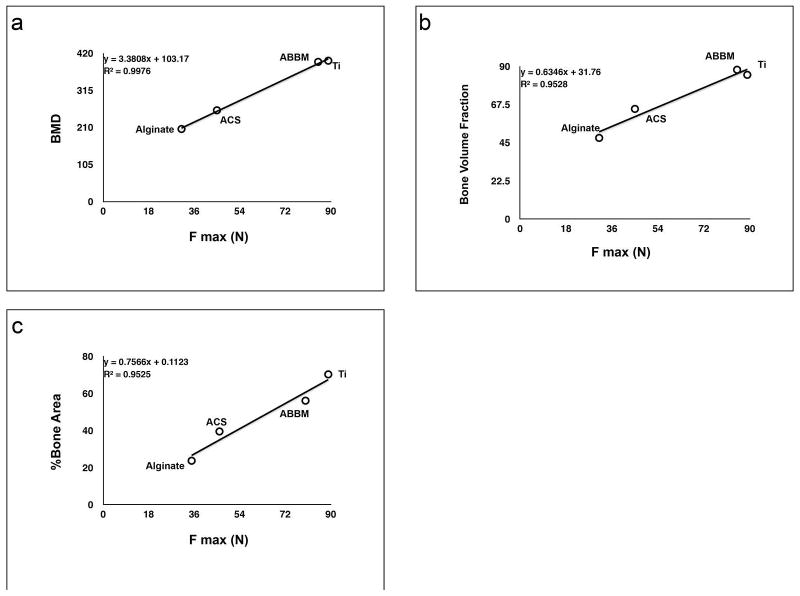
Recently we have demonstrated the ability of monoclonal antibodies (mAb) specific for bone morphogenetic protein (BMP)-2 immobilized on different scaffolds to mediate bone formation, a process referred to as Antibody Mediated Osseous Regeneration (AMOR). One of the key properties of regenerated bone is its biomechanical strength, in particular in load-bearing areas. This study sought to test the hypothesis that the biomechanical strength of regenerated bone depends of the mode of regeneration, as well as the scaffold used. Four different scaffolds, namely titanium granules (Ti), alginate hydrogel, anorganic bovine bone mineral (ABBM), and absorbable collagen sponge (ACS) were functionalized with anti-BMP-2 or isotype control mAb and implanted into rat critical-size calvarial defects. The morphology, density and strength of the regenerated bone were evaluated after 8 weeks. Results demonstrated that scaffolds functionalized with anti-BMP-2 mAb exhibited varying degrees of bone volume and density. Ti and ABBM achieved the highest bone volume, density, and strength of bone. When anti-BMP-2 mAb was immobilized on Ti or ABBM, the strength of the regenerated bone were 80% and 77% of native bone respectively, compared with 60% of native bone in sites implanted with rh-BMP-2. Control interventions with isotype mAb did not promote considerable bone regeneration and exhibited significantly lower mechanical properties. SEM analysis showed specimens immobilized with anti-BMP-2 mAb formed new bone with organized structure bridging the crack areas. Altogether, the present data demonstrated that the morphological and mechanical properties of bone bioengineered through AMOR could approximate that of native bone, when appropriate scaffolds are used. © 2015 Wiley Periodicals, Inc. J Biomed Mater Res Part B: Appl Biomater, 104B: 1465-1473, 2016.
Keywords: antibody-mediated bone regeneration; biomaterials; bone morphogenetic protein; mechanical properties; monoclonal antibody; tissue engineering.
© 2015 Wiley Periodicals, Inc.
Conflict of interest statement
The authors declare no potential conflicts of interest with respect to the authorship and/or publication of this article.
Figures


References
-
- Porter JR, Ruckh TT, Popat KC. Bone tissue engineering: a review in bone biomimetics and drug delivery strategies. Biotechnol Prog. 2009;25:1539–60. - PubMed
-
- Betz VM, Betz OB, Harris MB, Vrahas MS, Evans CH. Bone tissue engineering and repair by gene therapy. Front Biosci. 2008;13:833–41. - PubMed
-
- Sachlos E, Czernuszka JT. Making tissue engineering scaffolds work. Review on the application of solid freeform fabrication technology to the production of tissue engineering scaffold. Eur Cell Mater. 2003;5:29–40. - PubMed
-
- Tonegawa S. Somatic generation of antibody diversity. Nature. 1983;302:575–581. - PubMed
-
- Lin NH, Gronthos S, Bartold PM. Stem cells and periodontal regeneration. Aust Dent J. 2008;53:108–21. - PubMed
MeSH terms
Substances
Grants and funding
LinkOut - more resources
Full Text Sources
Other Literature Sources
Medical
