. 2015 Oct;14(10):1017-42.
doi: 10.1128/EC.00106-15. Epub 2015 Aug 7.

Eisosome Ultrastructure and Evolution in Fungi, Microalgae, and Lichens

Affiliations

Eisosome Ultrastructure and Evolution in Fungi, Microalgae, and Lichens

Jae-Hyeok Lee et al. Eukaryot Cell. 2015 Oct.

Abstract

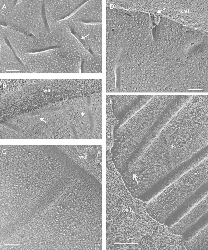
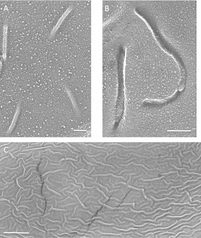
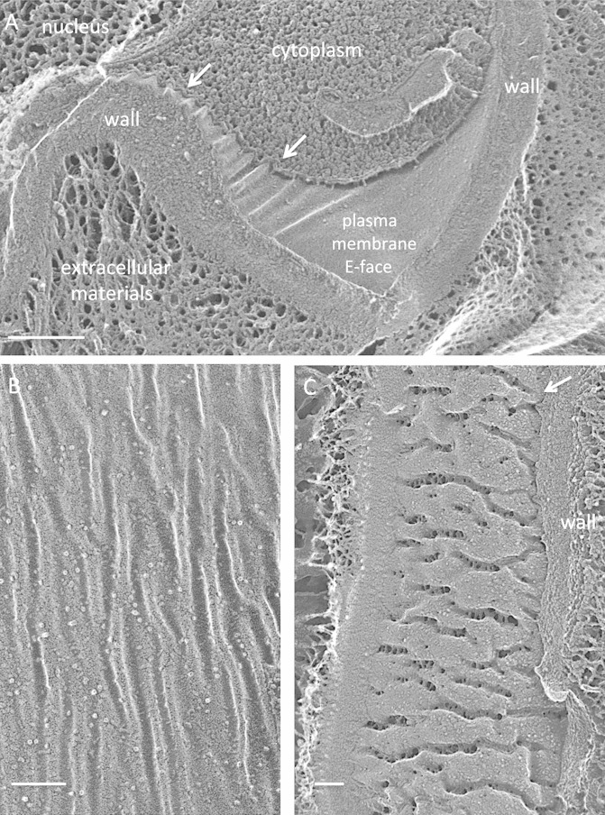
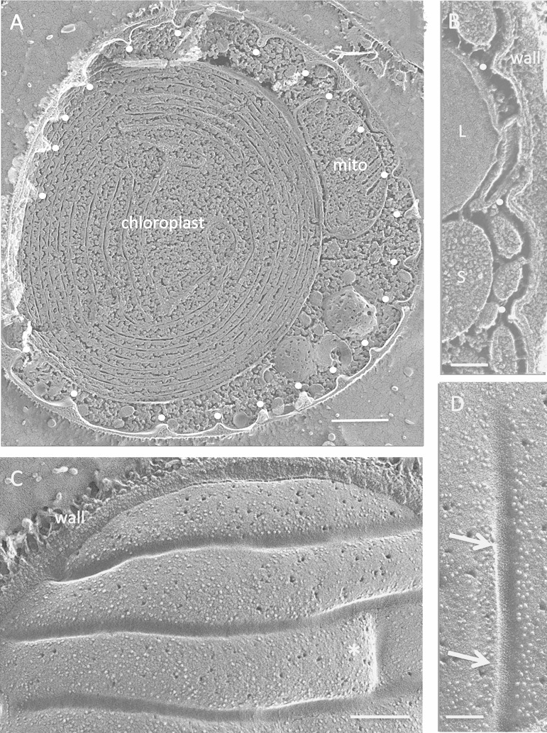
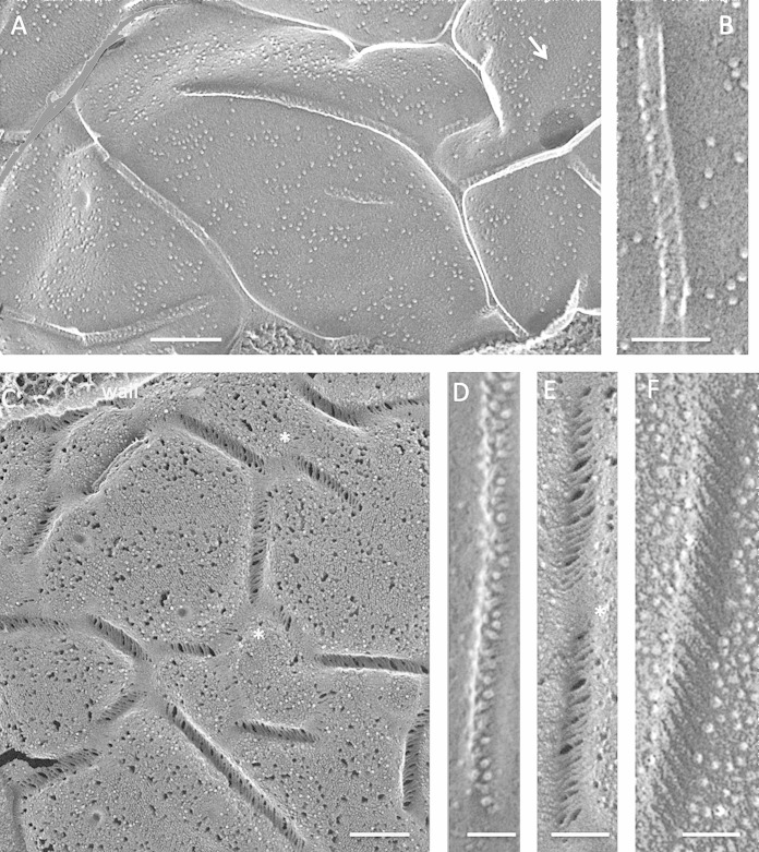
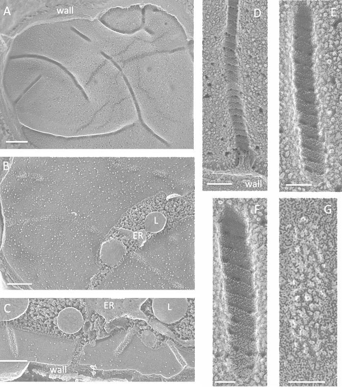
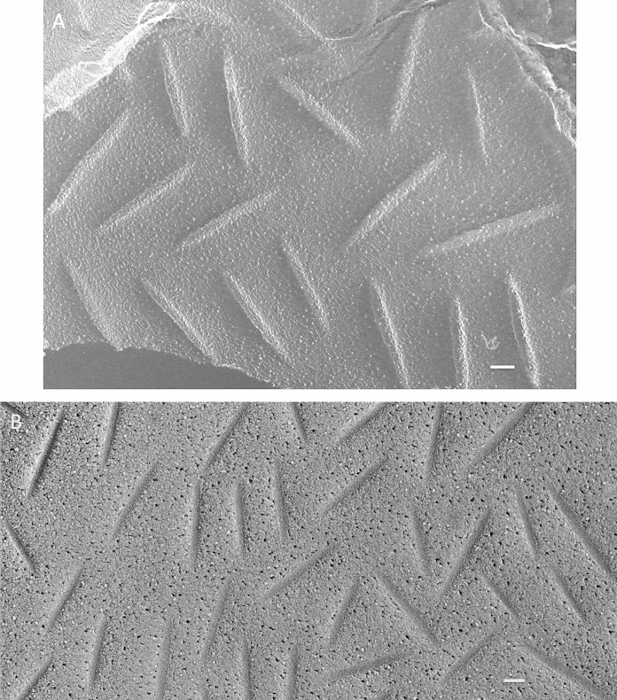
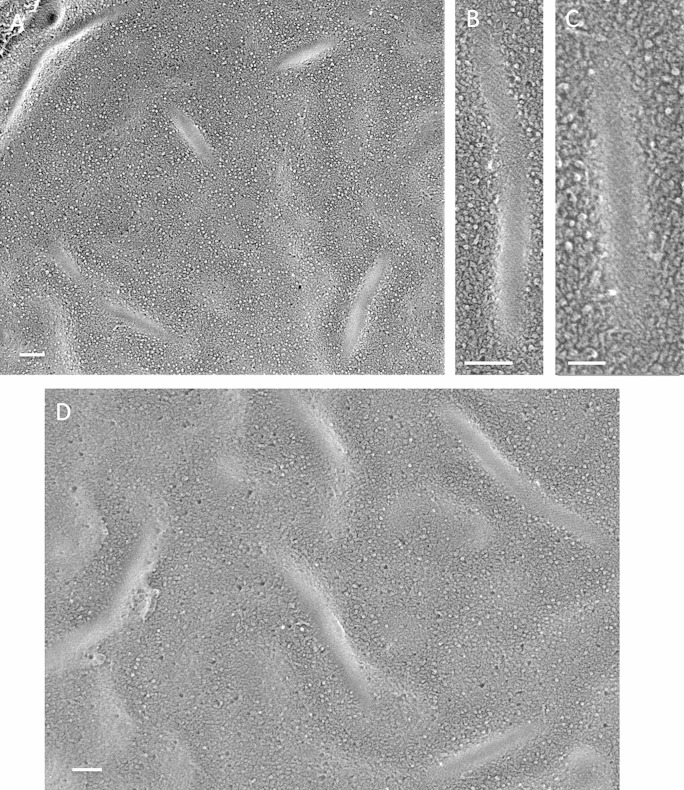
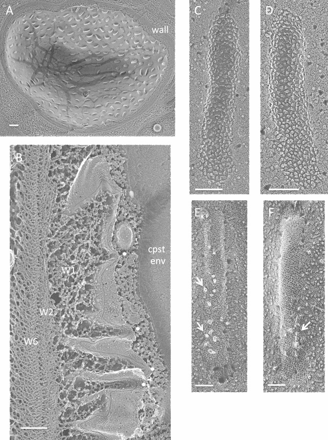
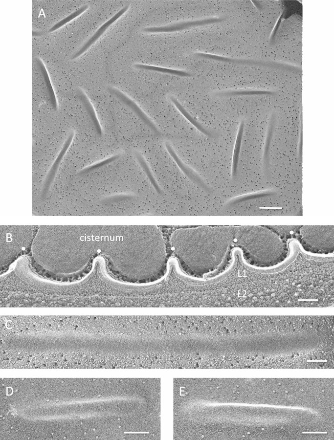
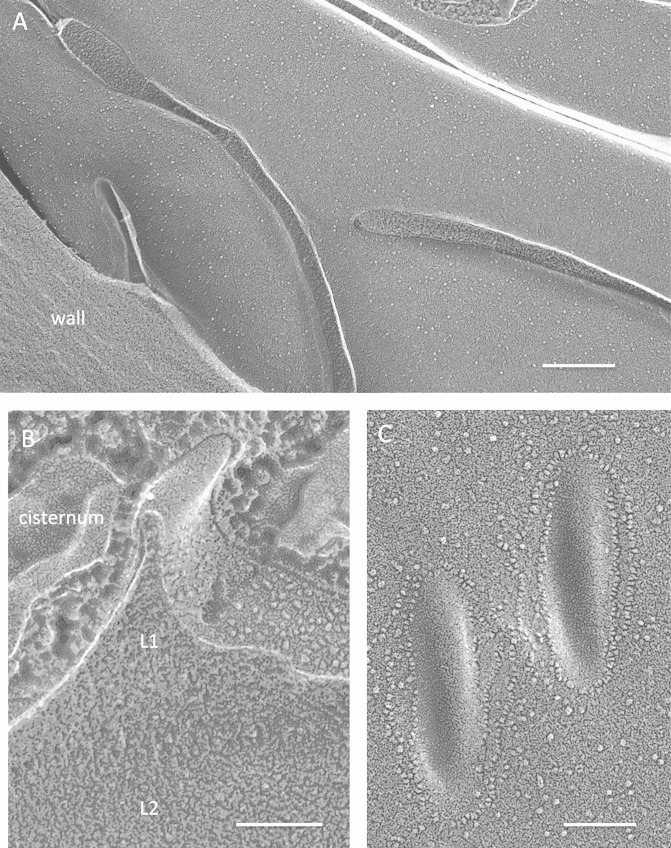
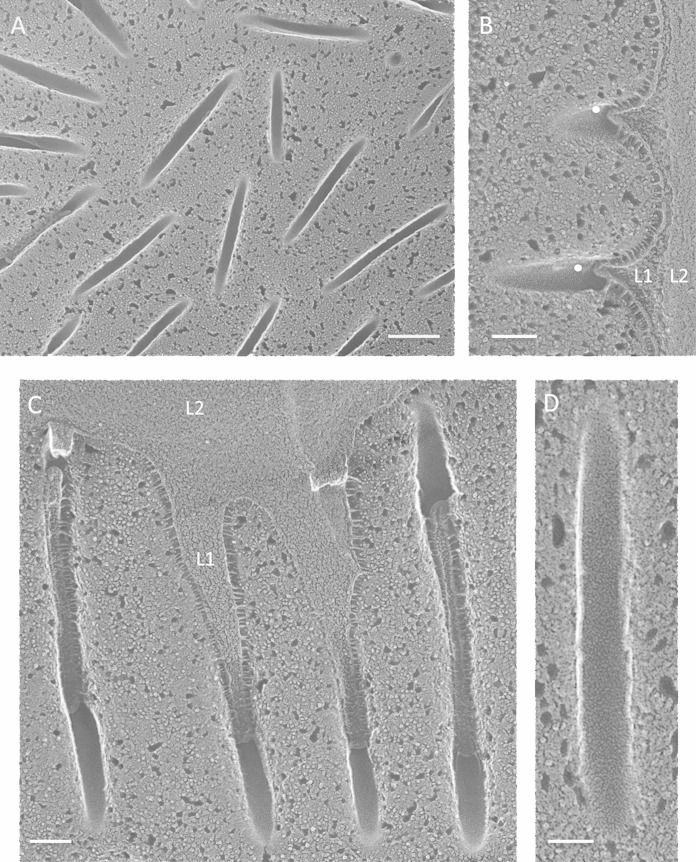
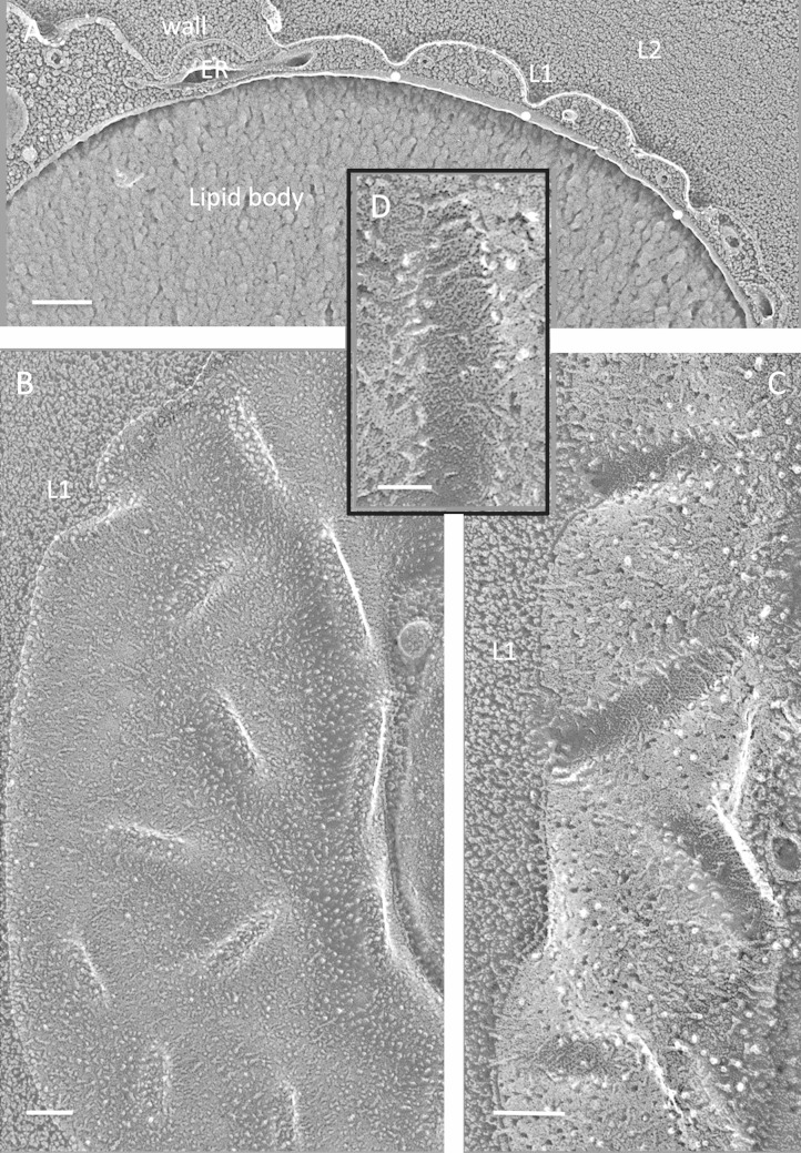
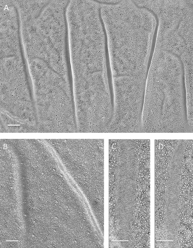
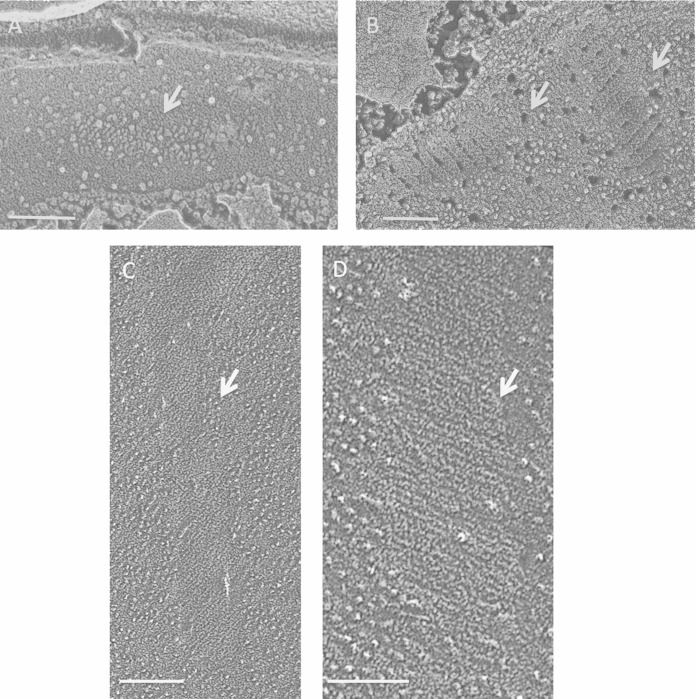
Eisosomes are among the few remaining eukaryotic cellular differentations that lack a defined function(s). These trough-shaped invaginations of the plasma membrane have largely been studied in Saccharomyces cerevisiae, in which their associated proteins, including two BAR domain proteins, have been identified, and homologues have been found throughout the fungal radiation. Using quick-freeze deep-etch electron microscopy to generate high-resolution replicas of membrane fracture faces without the use of chemical fixation, we report that eisosomes are also present in a subset of red and green microalgae as well as in the cysts of the ciliate Euplotes. Eisosome assembly is closely correlated with both the presence and the nature of cell walls. Microalgal eisosomes vary extensively in topology and internal organization. Unlike fungi, their convex fracture faces can carry lineage-specific arrays of intramembranous particles, and their concave fracture faces usually display fine striations, also seen in fungi, that are pitched at lineage-specific angles and, in some cases, adopt a broad-banded patterning. The conserved genes that encode fungal eisosome-associated proteins are not found in sequenced algal genomes, but we identified genes encoding two algal lineage-specific families of predicted BAR domain proteins, called Green-BAR and Red-BAR, that are candidate eisosome organizers. We propose a model for eisosome formation wherein (i) positively charged recognition patches first establish contact with target membrane regions and (ii) a (partial) unwinding of the coiled-coil conformation of the BAR domains then allows interactions between the hydrophobic faces of their amphipathic helices and the lipid phase of the inner membrane leaflet, generating the striated patterns.


Figures

FIG 1
FIG 1
Fungi. (A) Punctate concave eisosomal faces, some containing wall remnants (arrow), from an unidentified ascomycete bread mold. Bar, 100 nm. (B) Saccharomyces cerevisiae punctate concave eisosomal faces. Arrow, diagonal striations; asterisk, cluster of Pma1p proton pumps in the membrane compartment occupied by Pma1p (MCP). Bar, 100 nm. (C) Cryptococcus neoformans elongated concave eisosomal face with a uniform granular texture. Bar, 50 nm. (D) Candida albicans punctate convex eisosomal faces, with the wall entering at the arrow. Bar, 100 nm. (E) Schizosaccharomyces pombe elongated concave eisosomal faces. Arrow, diagonal striations; asterisk, eisosome with reduced depth and at right angles to others. Bar, 100 nm.
FIG 2
FIG 2
Lichenized Candelaria concolor mycobiont. (A) Sparse, straight, punctate convex faces. Bar, 100 nm. (B) Sparse, curved, punctate convex faces. Bar, 100 nm. (C) High-density elongated sinuous convex faces. Bar, 500 nm.
FIG 3
FIG 3
Nonlichenized Cladonia grayi (solo). (A) Hyphal eisosomes in both cross-fracture and planar fracture convex views, showing a concentration of eisosomes in the region of wall curvature (arrows) and their absence in the relatively flat domain. Bar, 200 nm. (B) High-density elongated sinuous convex faces. Bar, 100 nm. (C) High-density elongated sinuous concave faces carrying struts alternating with gaps. Arrow, wall entering eisosome. Bar, 100 nm.
FIG 4
FIG 4
Cyanidioschizon YNP 1A. (A) Cell survey with indented eisosome cross-fractures marked with white dots. Bar, 500 nm. (B) Eisosome indentations (white dots). L, lipid body; S, starch. Bar, 100 nm. (C) Elongated parallel eisosomes with a cross-segment at the asterisk (field enlarged from Fig. S6B in the supplemental material). Bar, 200 nm. (D) Concave face with regions of angled striations (arrows). Bar, 100 nm.
FIG 5
FIG 5
Galdieria sulfuraria. (A) Elongated anastomosing eisosomes, convex face. Arrow, rectangular membrane feature (see Fig. S10 in the supplemental material). Bar, 250 nm. (B) Convex face with few IMPs, displaying a diagonal pitch. Bar, 100 nm. (C) Long anastomosing concave eisosomes. Asterisks, domains lacking bands. Bar, 200 nm. (D) Convex face festooned with IMPs. Bar, 50 nm. (E) Concave face, banded. Bar, 100 nm. (F) Concave face, nonetched, banded. Bar, 50 nm.
FIG 6
FIG 6
Auxenochlorella protothecoides. (A) Elongated eisosomes that occasionally intersect, concave face. Bar, 250 nm. (B) Elongated parallel eisosomes, convex face. L, lipid body in cytoplasm. Bar, 200 nm. (C) Convex face. Bar, 200 nm. (D) Concave face. Bar, 100 nm. (E) Concave face. Bar, 50 nm. (F) Concave face. Bar, 50 nm. (G) Convex face. Bar, 50 nm.
FIG 7
FIG 7
Trebouxia jamesii. (A) High-density punctate eisosomes, convex face. Bar, 100 nm. (B) Concave face. Bar, 100 nm.
FIG 8
FIG 8
Asterochloris sp. (A) Punctate concave eisosomal faces. Bar, 100 nm. (B) Concave face with diagonal striations. Bar, 100 nm. (C) Concave face with diagonal striations. Bar, 50 nm. (D) Sinuous concave faces. Bar, 100 nm.
FIG 9
FIG 9
Chlamydomonas monoica. (A) Cell survey showing high-density punctate eisosomal topology. Bar, 500 nm. (B) The trabecular W1 layer of the wall associates with eisosomal invaginations (white dots); the dense W2 to W6 layers do not. Cpst env, chloroplast envelope. Bar, 250 nm. (C and D) Concave faces. Bars, 100 nm. (E and F) Convex faces. Arrows, irregularly shaped and distributed particles. Bars, 50 nm.
FIG 10
FIG 10
Chlamydomonas reinhardtii zygotes. (A) High-density punctate eisosomes, concave faces. Bar, 250 nm. (B) Eisosome cross-fractures (white dots). L1, penetrant granular wall layer; L2, denser wall layer. Bar, 100 nm. (C) Concave face. Bar, 100 nm. (D and E) Convex faces. Bars, 100 nm.
FIG 11
FIG 11
Polytomella parva cysts. (A) Elongated parallel eisosomes. Bar, 200 nm. (B) Eisosome cross-fracture (white dot). L1, granular wall layer; L2, denser wall layer. Bar, 100 nm. (C) Convex faces of punctate eisosomes encircled with chains of IMPs. Bar, 100 nm.
FIG 12
FIG 12
Haematococcus sp. cysts. (A) High-density punctate eisosomes, concave faces. Bar, 200 nm. (B) Cross-fractured eisosomes (white dots). L1, penetrant granular wall layer; L2, denser wall layer. Bar, 100 nm. (C) Parallel invaginations filled with L1 wall material, concave faces. Bar, 100 nm. (D) Concave face. Bar, 50 nm.
FIG 13
FIG 13
Borodinellopsis texensis cysts. (A) Cross-fractured eisosomes (white dots) containing the L1 but not the L2 wall layer. Bar, 200 nm. (B) Punctate convex faces. Bar, 100 nm. (C) Concave faces. Bar, 100 nm. (D) Enlargement of eisosome denoted with asterisk in panel C. Bar, 50 nm.
FIG 14
FIG 14
Euplotes cysts. (A) Elongated parallel eisosomes, convex faces. Bar, 100 nm. (B) Smooth convex faces. Bar, 50 nm. (C and D) Concave faces with diagonal striations. Bar, 50 nm.
FIG 15
FIG 15
Flat eisosomes (arrows). (A) Flat eisosome comparable to the convex face in Auxenochlorella protothecoides (c.f. Fig. 6C and G). Bar, 100 nm. (B) Flat eisosome comparable to the concave face in A. protothecoides (c.f. Fig. 6E and F). Bar, 100 nm. (C) Flat eisosome comparable to the convex face in Chlamydomonas monoica (c.f. Fig. 9E and F). Bar, 100 nm. (D) Flat eisosome comparable to the convex face in C. monoica (c.f. Fig. 9E and F). Bar, 100 nm.

References

    1. Moor H, Mühlethaler K. 1963. Fine structure in frozen-etched yeast cells. J Cell Biol 17:609–628. doi:10.1083/jcb.17.3.609. - DOI - PMC - PubMed
    1. Gross H, Kuebler O, Bas E, Moor H. 1978. Decoration of specific sites on freeze-fractured membranes. J Cell Biol 79:646–656. doi:10.1083/jcb.79.3.646. - DOI - PMC - PubMed
    1. Clarke KJ, Leeson EA. 1985. Plasmalemma structure in freezing tolerant unicellular algae. Protoplasma 129:120–126. doi:10.1007/BF01279909. - DOI
    1. Den Hertog AL, van Marle J, van Veen HA, Van't Hof W, Bolscher JGM, Veerman ECI, Niew Amerongen AV. 2005. Candidacidal effects of two antimicrobial peptides: histatin 5 causes small membrane defects, but LL-37 causes massive disruption of the cell membrane. Biochem J 388:689–695. doi:10.1042/BJ20042099. - DOI - PMC - PubMed
    1. Strádalová V, Stahlschmidt W, Grossman G, Blažĺková M, Rachel R, Tanner W, Malinsky J. 2009. Furrow-like invaginations of the yeast plasma membrane correspond to membrane compartment of Can1. J Cell Sci 122:2887–2894. doi:10.1242/jcs.051227. - DOI - PubMed

Publication types

MeSH terms

Substances

LinkOut - more resources