. 2015 Sep 1;142(17):2996-3008.
doi: 10.1242/dev.124586. Epub 2015 Aug 7.

Kinesin-1 interacts with Bucky ball to form germ cells and is required to pattern the zebrafish body axis

Affiliations

Kinesin-1 interacts with Bucky ball to form germ cells and is required to pattern the zebrafish body axis

Philip D Campbell et al. Development. .

Abstract

In animals, specification of the primordial germ cells (PGCs), the stem cells of the germ line, is required to transmit genetic information from one generation to the next. Bucky ball (Buc) is essential for germ plasm (GP) assembly in oocytes, and its overexpression results in excess PGCs in zebrafish embryos. However, the mechanistic basis for the excess PGCs in response to Buc overexpression, and whether endogenous Buc functions during embryogenesis, are unknown. Here, we show that endogenous Buc, like GP and overexpressed Buc-GFP, accumulates at embryonic cleavage furrows. Furthermore, we show that the maternally expressed zebrafish Kinesin-1 Kif5Ba is a binding partner of Buc and that maternal kif5Ba (Mkif5Ba) plays an essential role in germline specification in vivo. Specifically, Mkif5Ba is required to recruit GP to cleavage furrows and thereby specifies PGCs. Moreover, Mkif5Ba is required to enrich Buc at cleavage furrows and for the ability of Buc to promote excess PGCs, providing mechanistic insight into how Buc functions to assemble embryonic GP. In addition, we show that Mkif5Ba is also essential for dorsoventral (DV) patterning. Specifically, Mkif5Ba promotes formation of the parallel vegetal microtubule array required to asymmetrically position dorsal determinants (DDs) towards the prospective dorsal side. Interestingly, whereas Syntabulin and wnt8a translocation depend on kif5Ba, grip2a translocation does not, providing evidence for two distinct mechanisms by which DDs might be asymmetrically distributed. These studies identify essential roles for maternal Kif5Ba in PGC specification and DV patterning, and provide mechanistic insight into Buc functions during early embryogenesis.

Keywords: Bucky ball; Dorsoventral patterning; Germ cell; Germ plasm; Kinesin-1; Maternal.


Figures

Fig. 1.
Fig. 1.
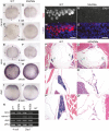
Endogenous Buc localizes to the germ plasm of embryos and binds Kinesin-1. (A,B) Endogenous Buc localizes to distal cleavage furrows of 4-cell embryos when chromosomes are decondensed (A), but not during metaphase (B) or anaphase. Insets show DAPI. Arrowheads indicate Buc accumulation. Scale bars: 100 µm for main images, 10 µm for insets. See text for quantification. (C) GFP-Buc, but not GFP, co-immunoprecipitates HA-Kif5Ba in HEK293T cells. (D-F) Overexpressed GFP-Buc localizes to cleavage furrows and overexpressed HA-Kif5Ba is expressed throughout blastomeres, potentially allowing them to interact in vivo. Arrowheads indicate Buc accumulation. (G) Schematic protein structure of Kif5Ba, illustrating motor, stalk and tail domains. Red dashed line indicates CRISPR target site. (H) DNA and predicted protein sequences of WT and mutant kif5Ba alleles (ae11 and ae12). Yellow indicates protospacer, orange indicates PAM, gray area/red dashed line indicates a deletion and pink indicates altered amino acids. (I) qRT-PCR for kif5Ba from pools of 5-dpf WT and homozygous mutants. kif5Ba undergoes nonsense-mediated decay in kif5Baae11/ae11 but not kif5Baae12/ae12 mutants. Error bars show mean±s.e.m.; Student's t-test, *P=0.0388.
Fig. 2.
Fig. 2.
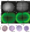
kif5Ba recruits germ plasm RNAs to cleavage furrows and promotes germ cell specification. (A,B,E,F) nanos3 (A,B) and vasa (E,F) localize to distal furrows in WT (A,E), but not in Mkif5Ba mutants (B,F), at 4-cell stage. Arrowheads indicate nanos3 and vasa accumulations. [nanos3: n=1/28 for Mkif5Baae11/ae12 (N=2 females), n=0/15 for Mkif5Baae11/ae11 (N=1 female), n=30/36 for WT (N=2 females) embryos have signal at ≥1 furrow; vasa: n=0/17 for Mkif5Baae11/ae12 (N=1 female), n=0/19 for Mkif5Baae11/ae11 (N=1 female), n=0/12 for Mkif5Baae12/ae12 (N=1 female), n=31/31 for WT (N=2 females) embryos have signal at ≥1 furrow.] (C,D,G,H) nanos3 (C,D) and vasa (G,H) are enriched in PGCs in WT (C,G), but not in Mkif5Ba mutants (D,H) at shield stage. Arrowheads indicate PGCs. [nanos3: n=0/32 for Mkif5Baae11/ae12 (N=2 females), n=0/19 for Mkif5Bae1/e1 (N=1 female), n=28/28 for WT (N=2 females) embryos have PGCs; vasa: n=0/18 for Mkif5Baae11/ae12 (N=1 female), n=0/22 for Mkif5Baae11/ae11 (N=1 female), n=0/17 for Mkif5Baae12/ae12 (N=1 female), n=37/37 for WT (N=2 females) embryos have PGCs.] (I,J) Vegetal localization of dazl is intact in WT (I) and Mkif5Ba mutants (J) at 4-cell stage. [n=19/19 for Mkif5Baae11/ae12 (N=2 females), n=20/20 for WT (N=2 females) embryos have vegetal signal.] (K) RT-PCR shows that germ plasm RNAs vasa, nanos3 and dazl are inherited by WT and Mkif5Ba mutant eggs (4-cell stage) and are maintained in WT and Mkif5Ba embryos (2 hpf). ef1α, loading control. NT, no template. (L-O) Vasa+ PGCs are present in WT (L,M) but not in Mkif5Ba mutant (N,O) embryos at 30 hpf. Scale bars: 10 µm. [n=0/48 for Mkif5Baae11/ae12 (N=2 females), n=19/67 for Mkif5Baae11/ae11 (N=3 females), n=0/20 for Mkif5Baae12/ae12 (N=3 females), n=53/53 for WT (N=4 females) have PGCs.] (P-Q″) H&E-stained transverse sections of adult WT (P) and MZkif5Ba mutants (Q) show that MZkif5Ba testes (green arrows) lack germ cells. P′,Q′ and P″,Q″ are higher magnification images of left and right testes, respectively. [n=4/4 Mkif5Baae12/ae12 adult males had empty testes; in addition, n=18/18 adult Mkif5Baae11/ae12 males dissected had testes devoid of germ cells.]
Fig. 3.
Fig. 3.
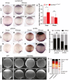
Overexpression of kif5Ba in Mkif5Ba mutants following fertilization is sufficient to specify germ cells. (A-C) nanos3 RNA is recruited to the first three cleavage furrows in WT (A) but is not recruited to furrows in Mkif5Ba mutants (B) at 16-cell stage. Injection of ha-kif5Ba restores nanos3 recruitment to 1-2 furrows in a fraction of Mkif5Ba mutants (C). Black arrowheads denote recruited nanos3. (D-F) nanos3 RNA becomes enriched in the prospective PGCs of WT (D) but is not enriched in Mkif5Ba mutants (E). Injection of ha-kif5Ba restores nanos3 enrichment to one PGC in a fraction of Mkif5Ba mutants (F). White arrowheads denote PGCs. (G-I) Vasa+ PGCs are present in WT (G) but not in Mkif5Ba mutants (H) at the yolk-yolk extension intersection. Injection of ha-kif5Ba restores small numbers of PGCs in Mkif5Ba mutants (arrowhead in I). See text for quantification. Scale bars: 50 µm.
Fig. 4.
Fig. 4.
Kif5Ba localizes Buc to cleavage furrows to mediate germ plasm assembly in the embryo. (A,B) GFP-Buc is recruited to distal furrows of WT (arrowheads in A) but not of Mkif5Ba mutants (B). [n=0/29 for Mkif5Baae11/ae12 (N=2 females), n=0/18 for Mkif5Baae11/ae11 (N=3 females), n=0/15 for kif5Baae12/ae12 (N=1 female), n=32/32 for WT (N=4 females) have compact distal GFP-Buc; n=2/29 for Mkif5Baae11/ae12, n=15/18 for Mkif5Baae11/ae11, n=0/15 for Mkif5Baae12/ae12 have some furrow recruitment of GFP-Buc, although distal furrow compaction is defective.] (C,D) Endogenous Buc localizes to distal cleavage furrows of WT (arrowheads in C) but not Mkif5Ba mutants (D). [n=2/15 for Mkif5Baae12/ae12 (N=2 females), n=2/17 for Mkif5Baae11/ae11 (N=1 female), n=24/32 for WT (N=4 females) of cells with decondensed chromosomes have Buc at ≥1 distal cleavage furrow.] (E-H) Compared with uninjected WT (E), gfp-buc injected WT (F) embryos have additional nanos3+ PGCs (arrowheads), whereas uninjected (G) and gfp-buc-injected (H) Mkif5Ba mutants lacked nanos3+ cells. See text for quantification.
Fig. 5.
Fig. 5.
Maternal kif5Ba is required for dorsal fate. (A-G) Mkif5Ba mutant embryos are variably ventralized at 1 dpf: V1, nearly complete AP axis lacking notochord and blocky somites (B); V2, reduced head structures (C); V3, largely lacking head structures except the posteriormost ones (D); V4, lack all head structures but possess an AP axis (E); V5, completely radialized axis (F). A small fraction of mutant embryos had duplicated anterior axes with fused caudal bodies (G,G′). (H) Distribution of phenotypic classes for homozygous and transheterozygous Mkif5Ba mutant embryos. The number of females assayed is indicated at the base of each bar. At least two clutches of embryos from each female were analyzed. Total number of embryos assayed for each genotype: n=1247 for Mkif5Baae11/ae12, n=2748 for Mkif5Baae11/ae11, n=2158 for Mkif5Baae12/ae12 and n=988 for siblings.
Fig. 6.
Fig. 6.
Maternal kif5Ba localizes dorsal determination components in activated eggs. (A-D′) sybu is vegetally localized at 2 mpa (A,B) and at 45 mpa (C,D) in both WT (A,C) and Mkif5Ba mutant (B,D) activated eggs. However, sybu is more dispersed at 2 mpa (B) and appears less abundant in most Mkif5Ba eggs at 45 mpa (D,D′). [2 mpa: n=30/30 for Mkif5Baae11/ae12 (N=2 females), n=29/29 for Mkif5Baae12/ae12 (N=1 female), n=63/63 for WT (N=3 females) have vegetally localized sybu; 45 mpa: quantification in panels.] (E) qRT-PCR reveals significantly more abundant sybu RNA in Mkif5Ba mutants at 2 mpa, although this RNA excess is not maintained at 45 mpa. Error bars show mean±s.d.; Student's t-test, *P=0.0219. (F-I) grip2a is vegetally localized at 2 mpa (F,G) and shifts asymmetrically by 45 mpa (H,I) in both WT (F,H) and Mkif5Ba mutant (G,I) activated eggs. [2 mpa: n=37/37 for Mkif5Baae11/ae12 (N=2 females), n=23/23 for WT (N=2 females) have vegetally localized grip2a; 45 mpa: see N for quantification.] (J-M) wnt8a is vegetally localized at 2 mpa in both WT (J) and Mkif5Ba mutant (K) activated eggs. However, wnt8a is asymmetric at 45 mpa in WT (L) but not Mkif5Ba mutant (M) activated eggs. [2 mpa: n=24/24 for Mkif5Baae11/ae12 (N=2 females), n=29/29 for WT (N=2 females) have vegetally localized wnt8a; 45 mpa: see N for quantification.] (N) Fraction of eggs exhibiting dorsally translocated RNA at 45 mpa. Number at base of columns indicates the number of eggs assayed. Chi-square, P=0.0312 (grip2a: n=2 females for each genotype; wnt8a: n=4 Mkif5Ba and n=3 WT females). (O-R″) Sybu is vegetally localized at 2 mpa (O) and is asymmetric at 45 mpa (P) in WT activated eggs. This is normal in some Mkif5Ba mutant eggs (Q,R), but others show aberrant localization at 2 mpa (Q′,Q″) and at 45 mpa (R′,R″). See text for quantification. (S) Injection of ha-kif5Ba RNA into 1-cell-stage Mkif5Ba mutants fails to suppress DV patterning defects. [n=138 uninjected and n=81 injected Mkif5Baae11/ae12 (N=1 female), n=172 uninjected and n=80 injected Mkif5Baae11/ae12 (N=2 females)]. Arrowheads designate the lateral limits of each expression domain.
Fig. 7.
Fig. 7.
Maternal kif5Ba organizes vegetal microtubules. (A-M) Vegetal microtubules are in a parallel orientation and bundled in WT activated eggs (A), but in Mkif5Ba mutants are either bundled and parallel (D), bundled but not parallel (G), or not bundled or parallel (J). Mkif5Ba mutant activated eggs display variable CG exocytosis defects (compare B with E,H,K). Importantly, the degree of CG retention does not correlate with the severity of the microtubule phenotype in Mkif5Ba mutants (M). Scale bars: 10 µm. Blue dotted line in M represents the upper limit of CG retention observed in WT activated eggs. Error bars show mean±s.d.; one-way ANOVA. (N-S) There is no difference between lateral microtubule organization in WT (N-P) and Mkif5Ba mutant (Q-S) activated eggs. Scale bars: 10 µm.
Fig. 8.
Fig. 8.
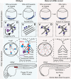
Model for Kif5Ba functions in dorsoventral patterning and germ cell formation. In WT activated eggs Kif5Ba mediates vegetal pMTA formation and dorsal determinant (DD) translocation towards the prospective dorsal side. Kif5Ba localizes endogenous Buc to distal cleavage furrows and promotes recruitment of GP-RNAs to furrows and subsequent PGC specification. In Mkif5Ba mutants the vegetal pMTA fails to form, resulting in ventralization. Furthermore, Buc localization to furrows is lost, resulting in failed GP recruitment to specify PGCs and adult sterility. Our data support a model whereby Kif5Ba is required to recruit Buc and GP-RNAs to cleavage furrows, although it is unclear whether it also acts to compact GP-RNAs within distal regions of the cleavage furrow.

Similar articles

Cited by

References

    1. Becker K. A. and Hart N. H. (1996). The cortical actin cytoskeleton of unactivated zebrafish eggs: spatial organization and distribution of filamentous actin, nonfilamentous actin, and myosin-II. Mol. Reprod. Dev. 43, 536-547. 10.1002/(SICI)1098-2795(199604)43:4<536::AID-MRD17>3.0.CO;2-X - DOI - PubMed
    1. Becker K. A. and Hart N. H. (1999). Reorganization of filamentous actin and myosin-II in zebrafish eggs correlates temporally and spatially with cortical granule exocytosis. J. Cell Sci. 112, 97-110. - PubMed
    1. Bontems F., Stein A., Marlow F., Lyautey J., Gupta T., Mullins M. C. and Dosch R. (2009). Bucky ball organizes germ plasm assembly in zebrafish. Curr. Biol. 19, 414-422. 10.1016/j.cub.2009.01.038 - DOI - PubMed
    1. Braat A. K., Zandbergen T., Van de Water S., Goos H. J. T. and Zivkovic D. (1999). Characterization of zebrafish primordial germ cells: morphology and early distribution of vasa RNA. Dev. Dyn. 216, 153-167. 10.1002/(SICI)1097-0177(199910)216:2<153::AID-DVDY6>3.0.CO;2-1 - DOI - PubMed
    1. Cai Q., Gerwin C. and Sheng Z.-H. (2005). Syntabulin-mediated anterograde transport of mitochondria along neuronal processes. J. Cell Biol. 170, 959-969. 10.1083/jcb.200506042 - DOI - PMC - PubMed

Publication types

MeSH terms