. 2015 Oct;8(5):1122-32.
doi: 10.1161/CIRCEP.115.002745. Epub 2015 Aug 7.

Identification and Functional Characterization of a Novel CACNA1C-Mediated Cardiac Disorder Characterized by Prolonged QT Intervals With Hypertrophic Cardiomyopathy, Congenital Heart Defects, and Sudden Cardiac Death

Affiliations

Identification and Functional Characterization of a Novel CACNA1C-Mediated Cardiac Disorder Characterized by Prolonged QT Intervals With Hypertrophic Cardiomyopathy, Congenital Heart Defects, and Sudden Cardiac Death

Nicole J Boczek et al. Circ Arrhythm Electrophysiol. 2015 Oct.

Abstract

Background: A portion of sudden cardiac deaths can be attributed to structural heart diseases, such as hypertrophic cardiomyopathy (HCM) or cardiac channelopathies such as long-QT syndrome (LQTS); however, the underlying molecular mechanisms are distinct. Here, we identify a novel CACNA1C missense mutation with mixed loss-of-function/gain-of-function responsible for a complex phenotype of LQTS, HCM, sudden cardiac death, and congenital heart defects.

Methods and results: Whole exome sequencing in combination with Ingenuity variant analysis was completed on 3 affected individuals and 1 unaffected individual from a large pedigree with concomitant LQTS, HCM, and congenital heart defects and identified a novel CACNA1C mutation, p.Arg518Cys, as the most likely candidate mutation. Mutational analysis of exon 12 of CACNA1C was completed on 5 additional patients with a similar phenotype of LQTS plus a personal or family history of HCM-like phenotypes and identified 2 additional pedigrees with mutations at the same position, p.Arg518Cys/His. Whole cell patch clamp technique was used to assess the electrophysiological effects of the identified mutations in CaV1.2 and revealed a complex phenotype, including loss of current density and inactivation in combination with increased window and late current.

Conclusions: Through whole exome sequencing and expanded cohort screening, we identified a novel genetic substrate p.Arg518Cys/His-CACNA1C, in patients with a complex phenotype including LQTS, HCM, and congenital heart defects annotated as cardiac-only Timothy syndrome. Our electrophysiological studies, identification of mutations at the same amino acid position in multiple pedigrees, and cosegregation with disease in these pedigrees provide evidence that p.Arg518Cys/His is the pathogenic substrate for the observed phenotype.

Keywords: Timothy syndrome; calcium channels, L-type; cardiomyopathy, hypertrophic; death, sudden, cardiac; genetics; long QT syndrome.


Figures

Figure 1
Figure 1
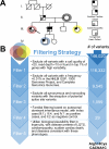
Pedigree and WES Filtering Strategy. (A) Pedigree with LQTS, SCD, cardiomyopathy, and CHDs (see key to right) that was utilized for WES. Individuals who underwent WES are highlighted with grey, red, yellow, and blue circles. (B) The filtering strategy utilized on this pedigree, showing the number of variants eliminated at each step.
Figure 2
Figure 2
Topology of CACNA1C. Shown is the topology of CACNA1C-encoded CaV1.2 in the membrane. The location of the p.Arg518Cys/His mutations, associated with cardiac-only Timothy syndrome, as identified in the pedigrees, are highlighted in red circles. In addition, other published CACNA1C mutations in Brugada syndrome (BrS)/Brugada syndrome + short QT syndrome (BrS+SQTS; black diamonds),, long QT syndrome (LQTS; black circles),, , - and Timothy syndrome (TS; white circles),, -, are highlighted. Shown in green is the AID (α-interaction domain; amino acids 428-445), the domain in which the β-subunit is known to bind. Variants with asterisks represent those that have been functionally characterized.
Figure 3
Figure 3
Pedigrees Harboring p.Arg518Cys/His. The original WES pedigree (pedigree 1), in addition to the pedigrees identified through the cohort analysis (pedigree 2 and 3) show the phenotypic status of each patient (key on right). In addition, each individual with a red circle is p.Arg518Cys/His positive, dotted red circles represent obligate positive p.Arg518Cys/His individuals, those who have been tested and are negative for p.Arg518Cys/His are demarcated with “neg”, and NA represents samples that were not available.
Figure 4
Figure 4
p.Arg518Cys/His Reduced ICaL Current Density in Heterologous Cells. (A) Whole cell ICaL current representative tracings from HEK293 cells expressing WT or mutant p.Arg518Cys/His determined from a holding potential of −90 mV to testing potential of +70 mV in 10 mV increments with 500 ms duration. (B) Current-voltage relationship for WT (n=15), and mutant p.Arg518Cys (n=14) and p.Arg518His (n=15) missense mutations. (C) Bar graph representing peak current density for WT (n=15), p.Arg518Cys (n=14), and p.Arg518His (n=15). All values represent mean ± SEM. *p<0.05 vs. CACNA1C-WT. (D) Confocal studies examining WT and mutant expression of N-terminally tagged CACNA1C with EYFP. The upper left corner of each image represents the DAPI nuclear stain, the upper right corner represents EYFP-CACNA1C, and the lower left corner shows the merged image. (E) Bar graph representing the peripheral:central fluorescence of WT (n=10) and p.Arg518Cys (n=10) cells. Bars represent mean ± SEM. *p<0.05 vs. CACNA1C-WT for Student's t-tests; *p<0.05 after ANOVA/Kruskal-Wallis with Dunn's correction for multiple comparisons.
Figure 5
Figure 5
CACNA1C-Arg518Cys/His Negatively Shifted ICaL V1/2 Inactivation. (A) Representative tracings of steady-state ICaL inactivation representing WT, p.Arg518Cys, and p.Arg518His determined from a holding potential of −90 mV to pre-pulse of 20 mV in 10 mV increments with 10 s duration followed by a test pulse of 30 mV with 500 ms duration. (B) Inactivation curves of ICaL WT (n=15), p.Arg518Cys (n=14), and p.Arg518His (n=15), determined from a holding potential of −90 mV to pre-pulse of 20 mV in 10 mV increments with 10 s duration followed by a test pulse of 30 mV with 500 ms duration. I/Imax represent normalized calcium current fitted with a Boltzmann function. Activation curves of ICaL WT (n=15), p.Arg518Cys (n=14), and p.Arg518His (n=15). G/Gmax represents normalized conductance fitted with a Boltzmann function. (C) Inactivation time constants (τ) for the fast phase of ICaL decay time of WT (n=11), p.Arg518Cys (n=10), and p.Arg518His (n=6) as a function of voltage. Time constants for each voltage step were determined by fitting a biexponential function to current decay. (D) Inactivation time constants (τ) for the slow phase of ICaL decay time of WT (n=11), p.Arg518Cys (n=10), and p.Arg518His (n=6) as a function of voltage. (E) Representative tracings of late current representing WT, p.Arg518Cys, and p.Arg518His measured at the end of 500 ms long depolarization of. (F) Normalized late current to peak current shown as percentages in a bar graph for WT, p.Arg518Cys, and p.Arg518His. All values represent mean ± SEM. *p<0.05 vs. CACNA1C-WT for Student's t-tests; *p<0.05 after ANOVA/Kruskal-Wallis with Dunn's correction for multiple comparisons.
Figure 6
Figure 6
Deceleration of VDI by p.Arg518Cys-CACNA1C. (A) Whole cell ICa and IBa current representative tracings from HEK293 cells expressing WT or mutant p.Arg518Cys determined from a holding potential of -90 mV to testing potential of +10 mV and +30 mV with 500 ms duration. (B) Voltage-dependent inactivation of ICa and IBa currents for WT and p.Arg518Cys channels (n=5 for each group, red *p<0.05 vs. WT IBa, black *p<0.05 vs. WT ICa; statisticscompleted for each of the 10 voltages). r500 represented fraction of current remaining after 500 ms depolarization normalized to peak current (r500).

References

    1. Go AS, Mozaffarian D, Roger VL, Benjamin EJ, Berry JD, Blaha MJ, Dai S, Ford ES, Fox CS, Franco S, Fullerton HJ, Gillespie C, Hailpern SM, Heit JA, Howard VJ, Huffman MD, Judd SE, Kissela BM, Kittner SJ, Lackland DT, Lichtman JH, Lisabeth LD, Mackey RH, Magid DJ, Marcus GM, Marelli A, Matchar DB, McGuire DK, Mohler ER, 3rd, Moy CS, Mussolino ME, Neumar RW, Nichol G, Pandey DK, Paynter NP, Reeves MJ, Sorlie PD, Stein J, Towfighi A, Turan TN, Virani SS, Wong ND, Woo D, Turner MB. Executive summary: Heart disease and stroke statistics--2014 update: A report from the American Heart Association. Circulation. 2014;129:399–410. - PubMed
    1. Chugh SS, Reinier K, Teodorescu C, Evanado A, Kehr E, Al Samara M, Mariani R, Gunson K, Jui J. Epidemiology of sudden cardiac death: clinical and research implications. Prog Cardiovasc Dis. 2008;51:213–228. - PMC - PubMed
    1. Boczek NJ, Tester DJ, Ackerman MJ. The molecular autopsy: An indispensable step following sudden cardiac death in the young? Herzschrittmacherth Elektrophysiol. 2012;23:167–173. - PMC - PubMed
    1. Maron BJ. Hypertrophic cardiomyopathy: a systematic review. JAMA. 2002;287:1308–1320. - PubMed
    1. Maron BJ, Towbin JA, Thiene G, Antzelevitch C, Corrado D, Arnett D, Moss AJ, Seidman CE, Young JB. Contemporary definitions and classification of the cardiomyopathies: An American Heart Association scientific statement from the Council on Clinical Cardiology, Heart Failure and Transplantation Committee; Quality of Care and Outcomes Research and Functional Genomics and Translational Biology Interdisciplinary Working Groups; and Council on Epidemiology and Prevention. Circulation. 2006;113:1807–1816. - PubMed

Publication types

Supplementary concepts