. 2015 Aug 1;29(15):1661-75.
doi: 10.1101/gad.265876.115.

Condensin promotes the juxtaposition of DNA flanking its loading site in Bacillus subtilis

Affiliations

Condensin promotes the juxtaposition of DNA flanking its loading site in Bacillus subtilis

Xindan Wang et al. Genes Dev. .

Abstract

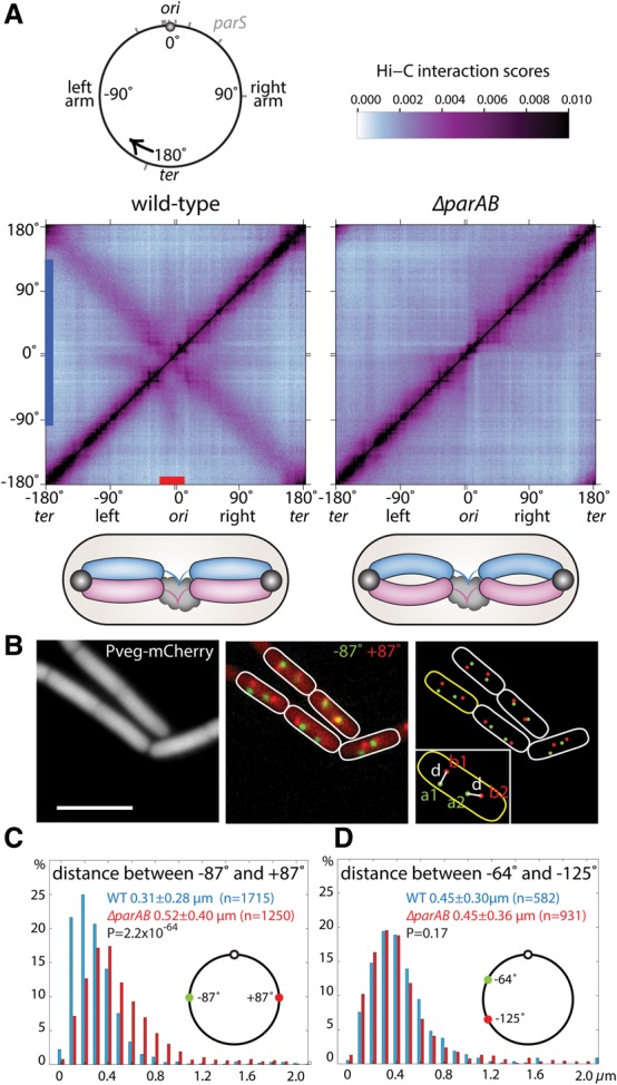
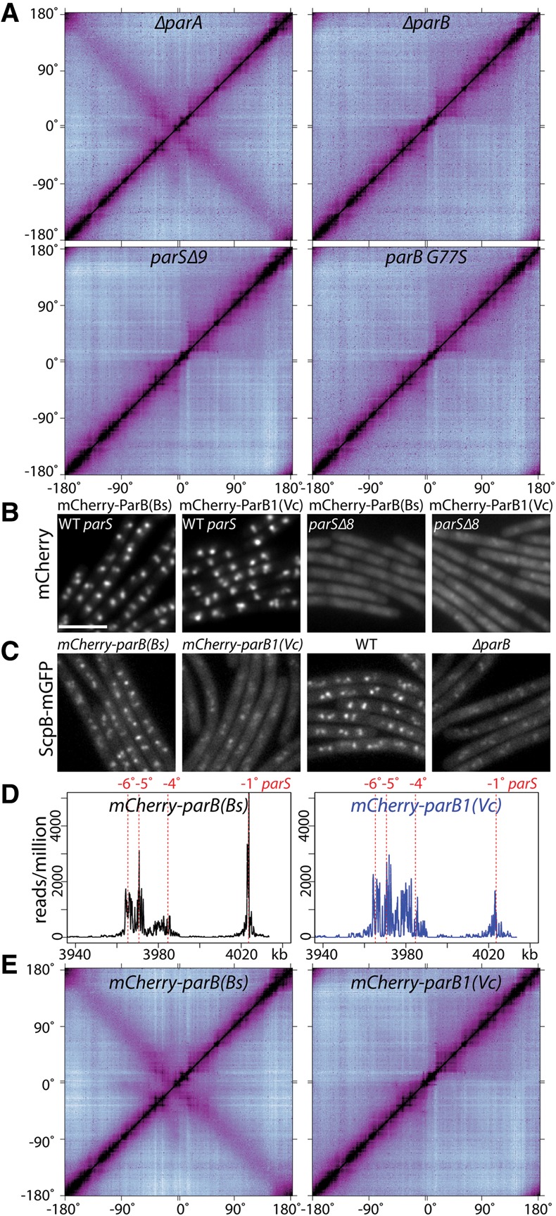
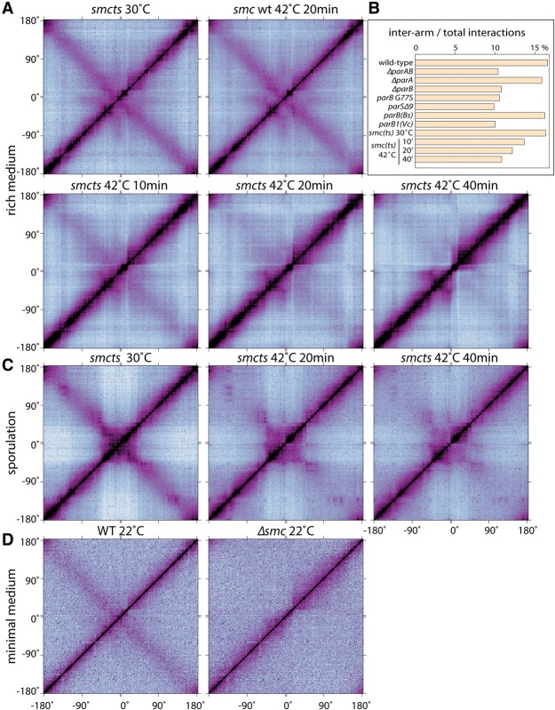
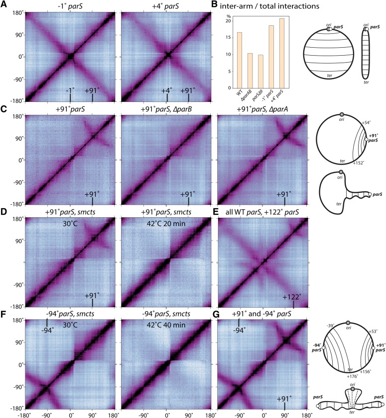
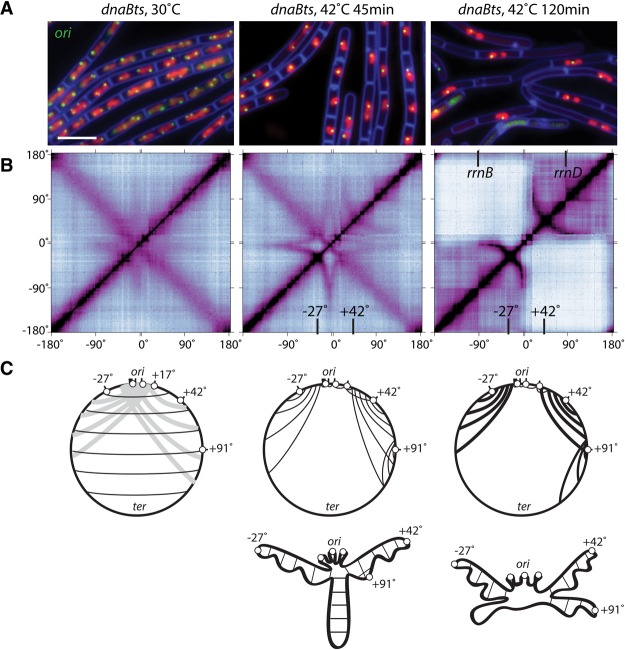
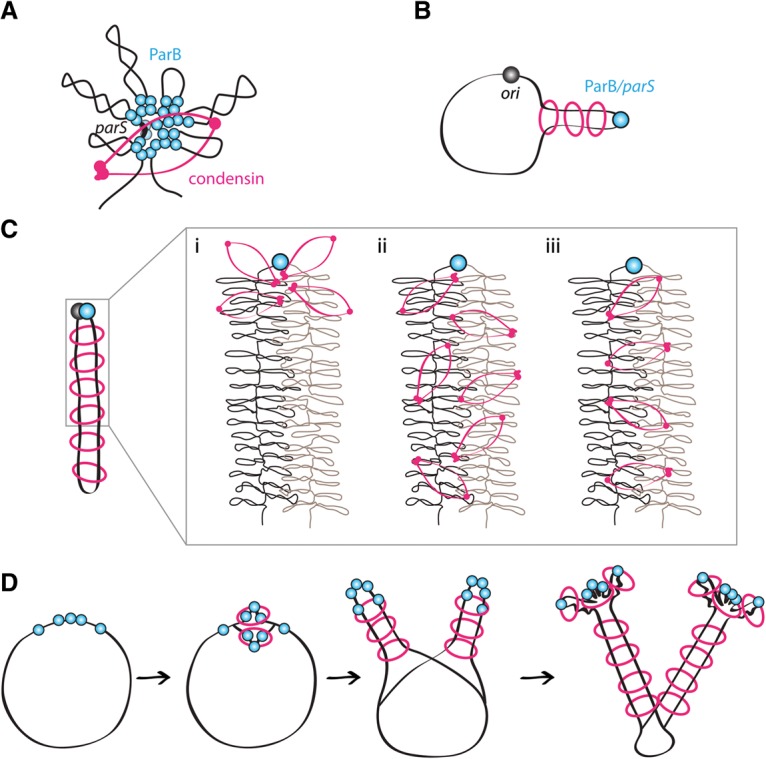
SMC condensin complexes play a central role in compacting and resolving replicated chromosomes in virtually all organisms, yet how they accomplish this remains elusive. In Bacillus subtilis, condensin is loaded at centromeric parS sites, where it encircles DNA and individualizes newly replicated origins. Using chromosome conformation capture and cytological assays, we show that condensin recruitment to origin-proximal parS sites is required for the juxtaposition of the two chromosome arms. Recruitment to ectopic parS sites promotes alignment of large tracks of DNA flanking these sites. Importantly, insertion of parS sites on opposing arms indicates that these "zip-up" interactions only occur between adjacent DNA segments. Collectively, our data suggest that condensin resolves replicated origins by promoting the juxtaposition of DNA flanking parS sites, drawing sister origins in on themselves and away from each other. These results are consistent with a model in which condensin encircles the DNA flanking its loading site and then slides down, tethering the two arms together. Lengthwise condensation via loop extrusion could provide a generalizable mechanism by which condensin complexes act dynamically to individualize origins in B. subtilis and, when loaded along eukaryotic chromosomes, resolve them during mitosis.

Keywords: DNA segregation; Hi-C; ParB; SMC; lengthwise condensation; mitosis; parS.


Figures

Figure 1.
Figure 1.
Long-range chromosome interactions require the parAB locus. (A) Normalized HindIII Hi-C interaction matrices displaying contact frequencies for pairs of 10-kb bins across the 4.0-Mb genome for wild-type (PY79) and ΔparAB (BDR1873) cells grown at 37°C in rich medium. Axes indicate the genome position of each bin in degrees. To more clearly visualize interactions in the origin region, the genome was oriented along the axes with the origin (ori) at the center and the left and right arms on either side (see Supplemental Fig. S1). An ∼300-kb region spanning the origin (red bar on the X-axis) and a broad region on the left and right arms (blue bar on the Y-axis) that have long-range interactions in wild-type are highlighted (see Supplemental Fig. S3A). The top left panel shows the circular B. subtilis chromosome with positions of the 10 parS sites (gray bars). The black arrow indicates the start and direction of the axes on the Hi-C contact maps. The top right panel has the color scale bar for all Hi-C contact maps presented in this study. Below the contact maps are schematics showing the ori-ter-ori configuration (Wang et al. 2014a) in B. subtilis cells and the presence or absence of interarm interactions in wild type and ΔparAB. Origins (black balls), chromosomal arms (blue and purple bars), and unreplicated DNA (black clouds) are shown. Long-range interactions emanating from the origin region are not shown. (B) Representative example of image analysis. Cytoplasmic mCherry was used to determine the outlines of the cells. Chromosomal loci at +87° and −87° were visualized using tetO/TetR-CFP and lacO/LacI-mYpet (BWX1912). Bar, 4 µm. (C) The distance between loci on opposing arms increases in cells lacking parAB (BWX3168) compared with wild type (BWX1912). (D) The distance between loci on the same arm remains unchanged in cells lacking parAB (BWX3446) compared with wild type (BWX3248). The X-axis is the interfocal distance in micrometers. The Y-axis is the percentage of cells that fall in each 0.1-µm bin. P-values were calculated using two-sample Kolmogorov-Smirnov test in Matlab. Details of analysis are in the Supplemental Material. Analyses of cells growing in minimal medium or cells lacking parA are in Supplemental Figure S3.
Figure 2.
Figure 2.
Long-range chromosome interactions require condensin recruitment to ParB/parS nucleoprotein complexes. (A) Normalized Hi-C contact matrices for cells lacking parA (BWX2876), parB (BDR2292), nine parS sites (parSΔ9; BWX3196), or a strain expressing ParB G77S (BDR2707) that binds parS but cannot form higher-order complexes. (BE) ParB1 from Vibrio cholerae [ParB1(Vc)] forms nucleoprotein complexes at parS sites in B. subtilis but does not recruit condensin or support long-range chromosomal interactions. (B) Localization of mCherry-ParB(Bs) and mCherry-ParB1(Vc) in cells with the full complement of parS sites (BWX3208 and BWX3209) or cells lacking eight origin-proximal parS sites (parSΔ8; BWX2519 and BWX2520). (C) Localization of ScpB-mGFP in cells expressing mCherry-ParB(Bs) (BWX2514), mCherry-ParB1(Vc) (BWX2515), wild-type ParB (BWX2030), or no ParB (ΔparB) (BWX2049). Fluorescence intensity was scaled to the same level for each channel. Bar, 4 µm. (D) Enrichment of mCherry-ParB(Bs) (BWX3208) and mCherry-ParB1(Vc) (BWX3209) as assayed by chromatin immunoprecipitation (ChIP) combined with deep sequencing (ChIP-seq) at a region of the B. subtilis chromosome (from 3940 to 4033 kb) encompassing four parS sites (at −1°, −4°, −5° and −6°). The number of reads per million reads was plotted on the Y-axis. mCherry-ParB(Bs) and mCherry-ParB1(Vc) spread to similar extents. ChIP-seq profiles for all of the individual parS sites and the entire genome are in Supplemental Figure S5. (E) Normalized Hi-C contact matrices of ΔparB mutants expressing mCherry-ParB(Bs) (BWX3208) or mCherry-ParB1(Vc) (BWX3209).
Figure 3.
Figure 3.
Condensin is required for long-range chromosome interactions. (A) Normalized Hi-C contact matrices of a temperature-sensitive smc mutant (smcts; BWX3151) grown at 30°C or after 10, 20, and 40 min at 42°C. A wild-type strain (smc WT; BWX2080) grown at 30°C and shifted for 20 min to 42°C is shown for comparison. (B) Bar graph of the percentage of interarm interactions compared with total interactions for the Hi-C experiments in A and Figures 1 and 2 (see Supplemental Fig. S6A for details). (C) The smcts strain (BWX3266) induced to sporulate at 30°C and harvested after 190 min at 30°C (left), 170 min at 30°C and then 20 min at 42°C (middle), or 150 min at 30°C and then 40 min at 42°C (right). (D) Wild-type (PY79) and Δsmc (BDR2298) strains grown in minimal medium at 22°C.
Figure 4.
Figure 4.
Condensin localizes along the chromosome arms during sporulation. (A) Representative fluorescence images of sporulating wild-type (BWX941) and ΔparAB (BWX3327) cells harboring an ScpB-YFP fusion. Cells were harvested 1 h after resuspension in sporulation medium. The replication origin was visualized using tetO/TetR-CFP, the membranes were stained with FM4-64, and the DNA was stained with DAPI. Bar, 4 µm. (B) Immunoblot analysis of the strains harboring ScpB-YFP in the presence (BKM1634) or absence (BWX3327) of ParAB. In the absence of ParAB, ScpB-YFP remained intact, and the level of SMC was similar to wild type. ScpB-YFP was detected using anti-GFP antibodies, and the arrowhead identifies the predicted size of free YFP. Both strains efficiently entered sporulation, as judged by the levels of the sporulation transcription factor SigF. SigA was used to control for loading.
Figure 5.
Figure 5.
Alignment of DNA flanking parS sites. (A) Normalized Hi-C contact matrices of strains with a single origin-proximal parS site at −1° (BDR2996) or +4° (BDR2985). Both strains also contain the weak parS site at +91°. A bar graph of the percentage of interarm interactions compared with total interactions for the indicated strains is shown in B (see Supplemental Fig. S6A for details). (C) Strains harboring the endogenous parS site at +91° but lacking all eight origin-proximal parS sites (parSΔ8) in an otherwise wild-type background (BNS1657) or in cells lacking parB (BWX2761) or parA (BDR3007). (D) A strain (BWX3304) harboring the endogenous parS site at +91° and smcts grown at 30°C and shifted for 20 min to 42°C. (E) A strain (BWX3172) with an ectopic parS site inserted at +122° in an otherwise wild-type background. (F) A strain (BWX3359) harboring an ectopic parS site at −94° and smcts grown at 30°C and shifted for 40 min to 42°C. A 20-min time point was not tested. (G) A strain (BWX3231) with two parS sites inserted to +91° and −94° in parSΔ9. The positions of the relevant parS sites (in degrees) are indicated in the contact maps. Schematic representations of the long-range interactions and the disposition of the chromosome are shown at the right (see also Supplemental Fig. S8A,B).
Figure 6.
Figure 6.
Long-range interactions emanating from the origin are revealed in cells blocked for replication initiation. (A) Representative fluorescence images of cells (BWX2533) harboring a temperature-sensitive replication initiation mutant in dnaB (dnaBts) at permissive (30°C) and restrictive (42°C) temperatures for the indicated times. The replication origins (ori) were visualized using tetO/TetR-CFP (green), the DNA was visualized with DAPI (red), and the cell membranes were visualized with FM4-64 (blue). Bar, 4 µm. (B) Normalized Hi-C contact matrices for the dnaBts strain (BNS1733) under the same conditions as in A. The locations of relevant parS sites at −27° and +42° and rrn loci are indicated (see also Supplemental Fig. S9). (C) Schematic representations of the long-range interactions and the disposition of the chromosome under the different conditions. The nine parS sites (black bars) are represented by six white balls: The cluster of −1°, −4°, −5°, and −6° parS sites are represented by a single ball, and the +4°, +17°, +42°, +91°, and −27° parS sites are represented by five separate balls. (Left panel) Light-gray lines in the schematic of cells grown under the permissive condition highlight the poorly resolved long-range interactions between the origin and more distal sites. (Middle panel) For simplicity, the interarm interactions are not shown, and only the zip-up interactions at the parS sites are presented. (Right panel) Thicker lines represent stronger interactions in the contact map.
Figure 7.
Figure 7.
Condensin rings tether DNA flanking parS sites and promote resolution of sister origins. (A) Schematic models of condensin (pink) loading at a ParB/parS nucleoprotein complex (light blue) (Graham et al. 2014; Wilhelm et al. 2015). (B) Condensin loaded at origin-distal parS sites aligns flanking DNA. (C) Condensin loaded at a single origin-proximal parS site juxtaposes the two arms. The insert shows three models for condensin action. (Panel i) Condensin acts adjacent to the parS site, setting up a compacted state that is propagated down the flanking DNA. The two condensed chromosomal arms are brought into close proximity by other proteins or due to confinement and/or crowding. (Panel ii) Condensin acts along the two arms to compact them and indirectly promotes their interaction. (Panel iii) Condensin loaded at parS encircles the flanking DNA strands. Migration down the DNA tethers the two strands of DNA. The supercoiled loops are shown schematically and are several times larger than depicted. (D) Condensin loading at newly replicated origin-proximal parS sites draws sister origins in on themselves and away from each other. Alignment of DNA flanking parS sites at −27° and +42° may impede movement of origin-loaded condensin rings down the arms. Although condensin rings are depicted encircling two strands of DNA in these models, the same activities would result from separate rings encircling DNA on either side of the loading site and migrating as unit (handcuffed) down the tethered DNA (see Supplemental Fig. S10).

References

    1. Alipour E, Marko JF. 2012. Self-organization of domain structures by DNA-loop-extruding enzymes. Nucleic Acids Res 40: 11202–11212. - PMC - PubMed
    1. Ben-Yehuda S, Rudner DZ, Losick R. 2003. RacA, a bacterial protein that anchors chromosomes to the cell poles. Science 299: 532–536. - PubMed
    1. Boonstra M, de Jong IG, Scholefield G, Murray H, Kuipers OP, Veening JW. 2013. Spo0A regulates chromosome copy number during sporulation by directly binding to the origin of replication in Bacillus subtilis. Mol Microbiol 87: 925–938. - PubMed
    1. Breier AM, Grossman AD. 2007. Whole-genome analysis of the chromosome partitioning and sporulation protein Spo0J (ParB) reveals spreading and origin-distal sites on the Bacillus subtilis chromosome. Mol Microbiol 64: 703–718. - PubMed
    1. Britton RA, Lin DC, Grossman AD. 1998. Characterization of a prokaryotic SMC protein involved in chromosome partitioning. Genes Dev 12: 1254–1259. - PMC - PubMed

Publication types

MeSH terms

Associated data