. 2015 Oct;4(10):1122-30.
doi: 10.5966/sctm.2015-0074. Epub 2015 Aug 7.

CXCL12 Gene Therapy Ameliorates Ischemia-Induced White Matter Injury in Mouse Brain

Affiliations

CXCL12 Gene Therapy Ameliorates Ischemia-Induced White Matter Injury in Mouse Brain

Yaning Li et al. Stem Cells Transl Med. 2015 Oct.

Abstract

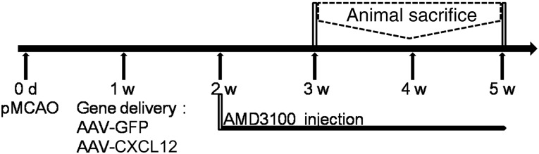
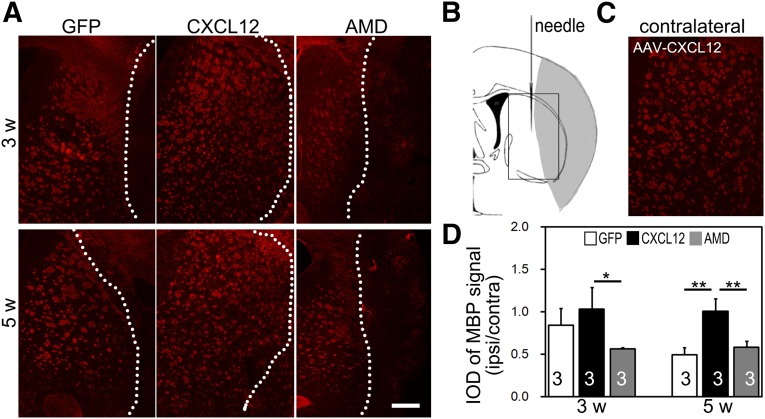
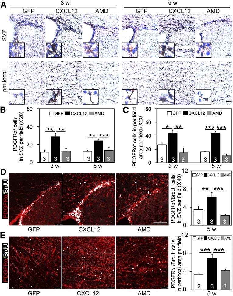
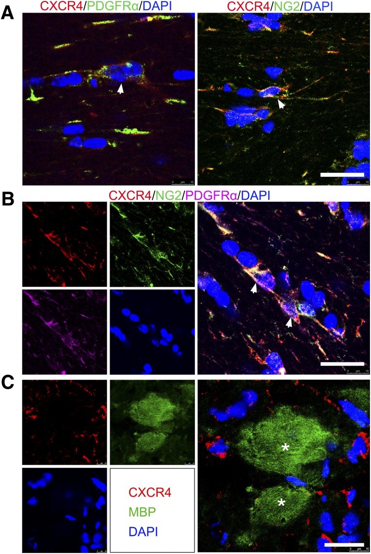
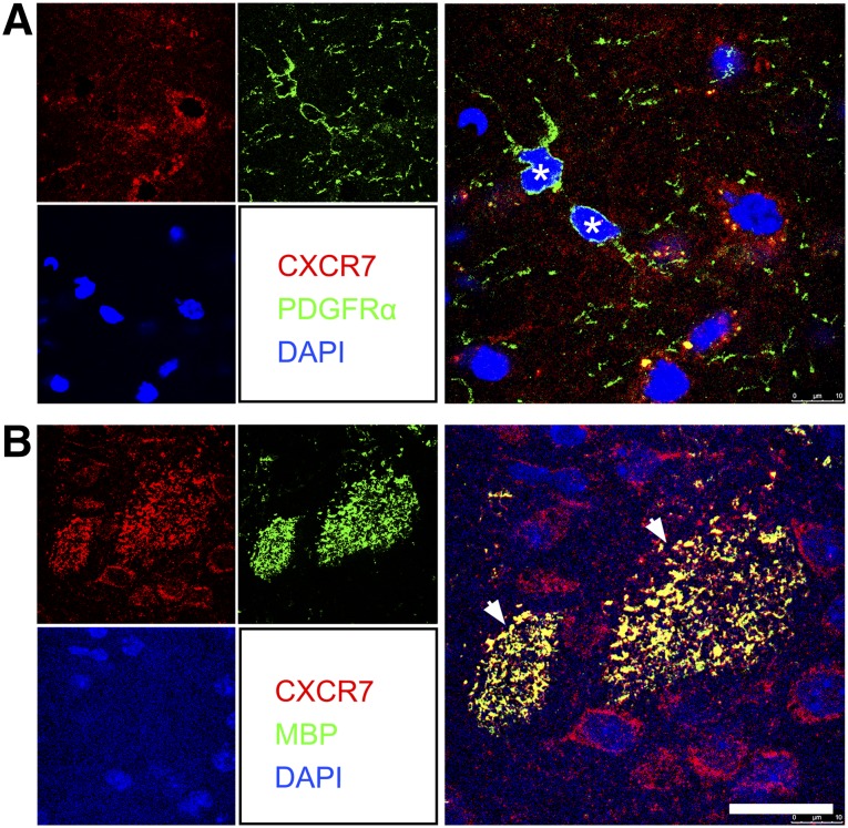
Remyelination is an important repair process after ischemic stroke-induced white matter injury. It often fails because of the insufficient recruitment of oligodendrocyte progenitor cells (OPCs) to the demyelinated site or the inefficient differentiation of OPCs to oligodendrocytes. We investigated whether CXCL12 gene therapy promoted remyelination after middle cerebral artery occlusion in adult mice. The results showed that CXCL12 gene therapy at 1 week after ischemia could protect myelin sheath integrity in the perifocal region, increase the number of platelet-derived growth factor receptor-α (PDGFRα)-positive and PDGFRα/bromodeoxyuridine-double positive OPCs in the subventricular zone, and further enhance their migration to the ischemic lesion area. Coadministration of AMD3100, the antagonist for CXCL12 receptor CXCR4, eliminated the beneficial effect of CXCL12 on myelin sheath integrity and negatively influenced OPC proliferation and migration. At 5 weeks after ischemia, CXCR4 was found on the PDGFRα- and/or neuron/glia type 2 (NG2)-positive OPCs but not on the myelin basic protein-positive mature myelin sheaths, and CXCR7 was only expressed on the mature myelin sheath in the ischemic mouse brain. Our data indicated that CXCL12 gene therapy effectively protected white matter and promoted its repair after ischemic injury. The treatment at 1 week after ischemia is effective, suggesting that this strategy has a longer therapeutic time window than the treatments currently available.

Significance: This study has demonstrated for the first time that CXCL12 gene therapy significantly ameliorates brain ischemia-induced white matter injury and promotes oligodendrocyte progenitor cell proliferation in the subventricular zone and migration to the perifocal area in the ischemic mouse brain. Additional data showed that CXCR4 receptor plays an important role during the proliferation and migration of oligodendrocyte progenitor cells, and CXCR7 might play a role during maturation. In contrast to many experimental studies that provide treatment before ischemic insult, CXCL12 gene therapy was performed 1 week after brain ischemia, which significantly prolonged the therapeutic time window of brain ischemia.

Keywords: C-X-C chemokine ligand 12; Ischemia; Oligodendrocyte progenitor cell; Remyelination; White matter.


Figures

Figure 1.
Figure 1.
Diagram of the experimental design. The mice underwent MCAO surgery at day 0 and received a stereotactic injection of AAV-CXCL12 or AAV-GFP (as control). AMD3100 was injected intraperitoneally from 2 weeks of ischemia. The mice were sacrificed at 3 and 5 weeks after ischemia. Abbreviations: AAV, adeno-associated virus; CXCL12, C-X-C chemokine ligand 12; d, day; GFP, green fluorescent protein; pMCAO, permanent middle cerebral artery occlusion; w, week.
Figure 2.
Figure 2.
Endogenous CXCL12 expression pattern in acute and postacute phases of ischemic mouse brain. (A): Confocal images of immunofluorescent double staining showed CXCL12 (red) expression in CD31+ (green) microvessels (Aa–Ac), NeuN+ (green) neurons (Ad–Af), GFAP+ (green) astrocytes (Aj–Al), and Iba1+ (green) microglials (Am–Ao) of sham mouse brain (column 1), 3-day postischemic mouse brain (column 2), and 14-day postischemic mouse brain (column 3). Lower magnification images of Iba1 immunostaining in the sham mouse brain (Ap) and 3 days (Aq) and 14 days (Ar) of ischemic mouse brain are also shown. (B): Enzyme-linked immunosorbent assay quantification of CXCL12 in the postischemic mouse brain at 1 hour and 1, 3, 7, and 14 days of MCAO (n = 3 per group; numbers indicated on each column). Scale bars = 20 μm (Ao), 100 μm (Ar). Data are presented as mean ± SD; ∗, p < .05; ∗∗, p < .01. Abbreviations: CXCL12, C-X-C chemokine ligand 12; d, day; DAPI, 4′,6-diamidino-2-phenylindole; GFAP, glial fibrillary acidic protein; h, hour; pMCAO, permanent middle cerebral artery occlusion.
Figure 3.
Figure 3.
Postacute CXCL12 gene therapy protects myelin sheath integrity. (A): Immunofluorescent staining of MBP+ myelin sheath in the perifocal region of the ipsilateral hemisphere. (B): Hollow box in schematic brain diagram shows the area of interest, the perifocal striatum in ischemic mice. (C): Immunofluorescent staining of MBP+ myelin sheath in the contralateral hemisphere of an AAV-CXCL12 transferred mouse. (D): IOD quantification of MBP+ signal after MCAO in AAV-GFP, AAV-CXCL12, and AAV-CXCL12/AMD3100 treated groups (n = 3 per group; numbers indicated in each column). Scale bar = 500 μm. Data are presented as mean ± SD. ∗, Statistical significance, p < .05; ∗∗, p < .01. Abbreviations: AMD, adeno-associated virus-C-X-C chemokine ligand 12-AMD3100; CXCL12, adeno-associated virus-C-X-C chemokine ligand 12; GFP, adeno-associated virus-green fluorescent protein; IOD, integral optical density; ipsi/contra, ipsilateral/contralateral; MBP, myelin basic protein; w, week.
Figure 4.
Figure 4.
Postacute CXCL12 gene therapy promotes OPC proliferation and migration in ischemic mice. (A): Representative photomicrographs of 3,3′-diaminobenzidine-stained coronal sections showing PDGFRα+ cells in SVZ (panel 1) and ipsilateral perifocal region (panel 2). Insets show higher magnifications from SVZ and perifocal region, respectively. Quantifications of PDGFRα+ cells in SVZ (B) and perifocal region (C) after middle cerebral artery occlusion (MCAO) in AAV-GFP, AAV-CXCL12, and AAV-CXCL12/AMD3100 treated groups (n = 3 per group; numbers indicated in each column). Double immunostaining of PDGFRα (red) and BrdU (white) positive cells in SVZ (D) and perifocal area (E) after 5 weeks of MCAO; quantification shown by each bar graph (n = 3 per group). Scale bars = 100 μm. Data are presented as mean ± SD; ∗, p < .05; ∗∗, p < .01; ∗∗∗, p < .001. Abbreviations: AMD, adeno-associated virus-C-X-C chemokine ligand 12-AMD3100; BrdU, bromodeoxyuridine; CXCL12, adeno-associated virus-C-X-C chemokine ligand 12; GFP, adeno-associated virus-green fluorescent protein; OPCs, oligodendrocyte progenitor cells; PDGFRα, platelet-derived growth factor receptor-α; SVZ, subventricular zone; w, week.
Figure 5.
Figure 5.
CXCR4 was expressed on OPCs but not on mature myelin sheaths in the 5-week postischemic mouse brain. (A): Confocal images showing CXCR4 (red) expressed on PDGFRα+ and NG2+ cells (green, arrows). (B): Confocal images of immunofluorescent triple staining of frozen coronal sections showing that CXCR4 (red) is coexpressed by cells that stained double positive for PDGFRα (purple, arrows) and NG2 (green, arrows). (C): CXCR4 (red) was not detected on MBP+ myelin sheath cells (green, asterisks). Scale bars = 20 μm. Abbreviations: CXCR, C-X-C receptor; DAPI, 4′,6-diamidino-2-phenylindole; MBP, myelin basic protein; OPCs, oligodendrocyte progenitor cells; PDGFRα, platelet-derived growth factor receptor-α.
Figure 6.
Figure 6.
CXCR7 was not detected on OPCs but on mature myelin sheaths in the 5-week postischemic mouse brain. Double immunostaining confocal images showed CXCR7 (red) was barely detected on PDGFRα+ cells (green, asterisks) (A) but coexpressed with MBP+ mature myelin sheath (green, arrows) (B). Scale bar = 20 μm. Abbreviations: CXCR, C-X-C receptor; DAPI, 4′,6-diamidino-2-phenylindole; MBP, myelin basic protein; OPCs, oligodendrocyte progenitor cells; PDGFRα, platelet-derived growth factor receptor-α.

References

    1. Pantoni L, Garcia JH, Gutierrez JA. Cerebral white matter is highly vulnerable to ischemia. Stroke. 1996;27:1641–1647. - PubMed
    1. Arai K, Lo EH. Experimental models for analysis of oligodendrocyte pathophysiology in stroke. Exp Transl Stroke Med. 2009;1:6. - PMC - PubMed
    1. Lo EH, Dalkara T, Moskowitz MA. Mechanisms, challenges and opportunities in stroke. Nat Rev Neurosci. 2003;4:399–415. - PubMed
    1. Fork M, Bartels C, Ebert AD, et al. Neuropsychological sequelae of diffuse traumatic brain injury. Brain Inj. 2005;19:101–108. - PubMed
    1. Irvine KA, Blakemore WF. Remyelination protects axons from demyelination-associated axon degeneration. Brain. 2008;131:1464–1477. - PubMed

Publication types

MeSH terms