Case Reports
. 2016 Jun;10(2):245-51.
doi: 10.1007/s12105-015-0643-z. Epub 2015 Aug 8.

Primary Xanthoma of the Mandible: Report of a Rare Case

Affiliations
Case Reports

Primary Xanthoma of the Mandible: Report of a Rare Case

Dominic Morel et al. Head Neck Pathol. 2016 Jun.

Abstract

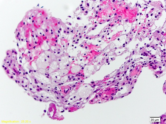
Xanthoma is a lesion most commonly seen in soft tissues such as the skin, subcutis, or tendon sheaths. Xanthoma formation is often associated with primary or secondary hyperlipidemia. Primary bone xanthomas are extremely rare benign bone lesions not associated with hyperlipidemia, histopathologically characterized by histiocytes, abundant lipid containing macrophages (foam cells), and multinucleated giant cells. Cholesterol clefts can be found in the medullary bone. Less than ten cases of xanthoma in the mandible have been reported. We present a rare primary intrabony xanthoma in a normolipidemic patient.

Keywords: Foamy histiocytes; Intraosseous xanthoma; Mandible; Non-Langerhans histiocytic process; Xanthoma.


Figures

Fig. 1
Fig. 1
Panoramic radiograph on presentation. Note the positioning error, depicted by larger teeth on the left side
Fig. 2
Fig. 2
CBCT on presentation. Axial (a), coronal (b), and 3D reconstruction (c) views
Fig. 3
Fig. 3
Photomicrographs. Low power (×10) H + E stain showing histiocytic cells involving and infiltrating trabecular bone in a background of loose fibrous connective tissue devoid of significant inflammatory infiltrate
Fig. 4
Fig. 4
High magnification (×40) H + E stain demonstrating the abundance of foamy histiocytes
Fig. 5
Fig. 5
Xanthoma cells exhibiting strong positive CD68 staining
Fig. 6
Fig. 6
Panoramic radiograph at 6 month follow-up

References

    1. Huang GS, Huang CW, Lee CW, et al. Xanthoma of the sacrum. Skeletal Radiol. 2004;33:674–678. doi: 10.1007/s00256-004-0829-z. - DOI - PubMed
    1. Muthusamy KA, Azmi K, Narayanan P, et al. Bilateral temporal bone xanthoma: case report. J Neurosurg. 2008;108:361–364. doi: 10.3171/JNS/2008/108/2/0361. - DOI - PubMed
    1. Marqués Mateo M, Puche Torres M, Miragall Alba L, et al. Primary mandibular bone xanthoma: a case report. Int J Oral Maxillofac Surg. 2004;33:86–87. doi: 10.1016/j.ijom.2003.12.006. - DOI - PubMed
    1. Harsanyi BB, Larsson A. Xanthomatous lesions of the mandible: osseous expression of non-X histiocytosis and benign fibrous histiocytoma. Oral Surg Oral Med Oral Pathol. 1988;65:551–566. doi: 10.1016/0030-4220(88)90138-7. - DOI - PubMed
    1. de Moraes Ramos-Perez FM, de Pádua JM, Silva-Sousa YTC, et al. Primary xanthoma of the mandible. Dentomaxillofac Radiol. 2011;40:393–396. doi: 10.1259/dmfr/51850495. - DOI - PMC - PubMed

Publication types

LinkOut - more resources