

Organotypic brain slice cultures: A review
- PMID: 26254240
- PMCID: PMC4699268
- DOI: 10.1016/j.neuroscience.2015.07.086
Organotypic brain slice cultures: A review
Abstract
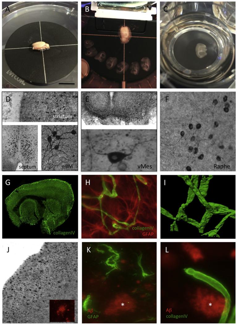
In vitro cell cultures are an important tool for obtaining insights into cellular processes in an isolated system and a supplement to in vivo animal experiments. While primary dissociated cultures permit a single homogeneous cell population to be studied, there is a clear need to explore the function of brain cells in a three-dimensional system where the main architecture of the cells is preserved. Thus, organotypic brain slice cultures have proven to be very useful in investigating cellular and molecular processes of the brain in vitro. This review summarizes (1) the historical development of organotypic brain slices focusing on the membrane technology, (2) methodological aspects regarding culturing procedures, age of donors or media, (3) whether the cholinergic neurons serve as a model of neurodegeneration in Alzheimer's disease, (4) or the nigrostriatal dopaminergic neurons as a model of Parkinson's disease and (5) how the vascular network can be studied, especially with regard to a synthetic blood-brain barrier. This review will also highlight some limits of the model and give an outlook on future applications.
Keywords: cholinergic; dopaminergic; organotypic; vascular; whole-brain cultures.
Copyright © 2015 The Author. Published by Elsevier Ltd.. All rights reserved.
Figures
References
-
- Alberdi E, Sánchez-Gómez MV, Cavaliere F, Pérez-Samartín A, Zugaza JL, Trullas R, Domercq M, Matute C. Amyloid beta oligomers induce Ca2+ dysregulation and neuronal death through activation of ionotropic glutamate receptors. Cell Calcium. 2010;47(3):264–272. - PubMed
-
- Allen YS, Devanathan PH, Owen GP. Neurotoxicity of beta-amyloid protein: cytochemical changes and apoptotic cell death investigated in organotypic cultures. Clin Exp Pharmacol Physiol. 1995;22:370–371. - PubMed
-
- Annis CM, Edmond J, Robertson RT. A chemically-defined medium for organotypic slice cultures. J Neurosci Methods. 1990;32:63–70. - PubMed
-
- Ansevin KD, Lipps BV. Tissue plate: a new technique for long-termorganotypic culture. In Vitro. 1973;8:483–488. - PubMed
Publication types
MeSH terms
Grants and funding
LinkOut - more resources
Full Text Sources
Other Literature Sources
