. 2015 Oct;309(8):H1271-80.
doi: 10.1152/ajpheart.00517.2014. Epub 2015 Aug 7.

ErbB2 overexpression upregulates antioxidant enzymes, reduces basal levels of reactive oxygen species, and protects against doxorubicin cardiotoxicity

Affiliations

ErbB2 overexpression upregulates antioxidant enzymes, reduces basal levels of reactive oxygen species, and protects against doxorubicin cardiotoxicity

Frances Belmonte et al. Am J Physiol Heart Circ Physiol. 2015 Oct.

Abstract

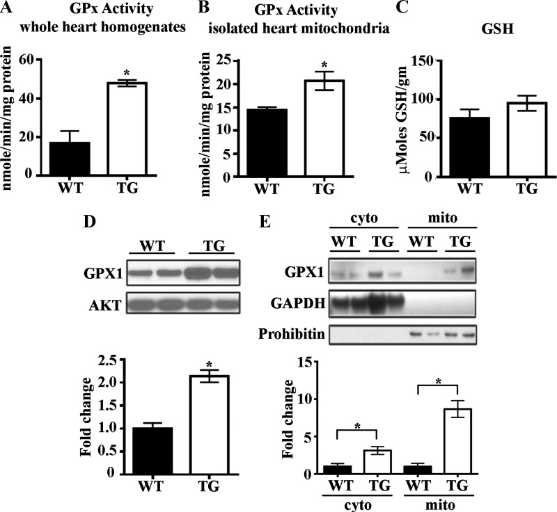
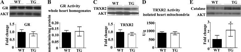
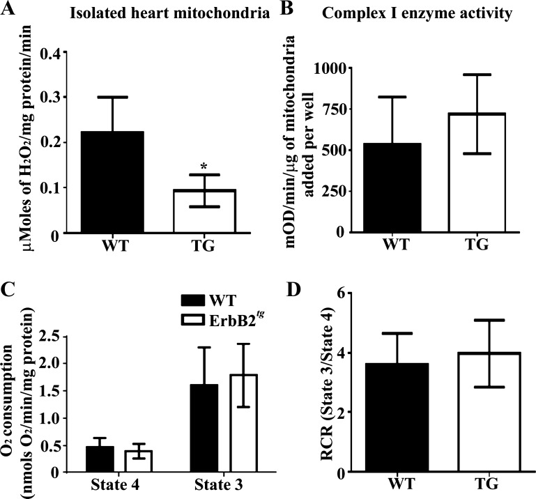
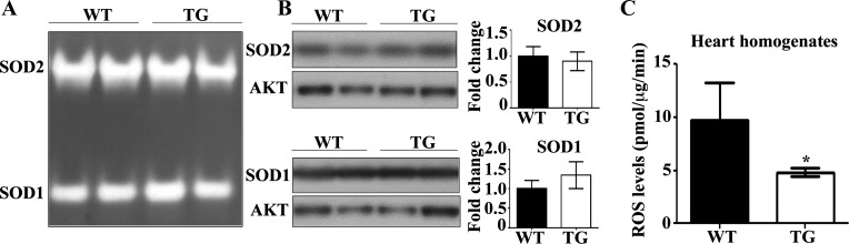
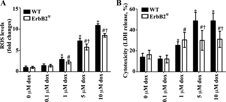
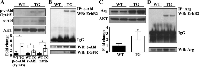
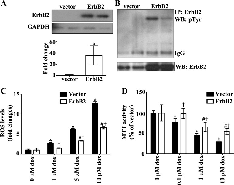
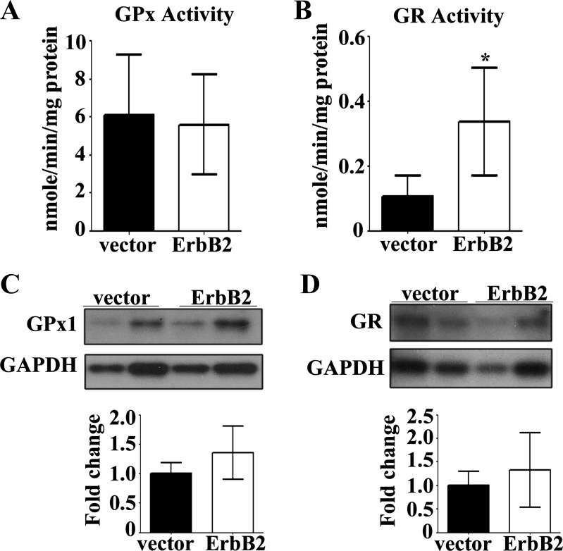
Levels of the HER2/ErbB2 protein in the heart are upregulated in some women during breast cancer therapy, and these women are at high risk for developing heart dysfunction after sequential treatment with anti-ErbB2/trastuzumab or doxorubicin. Doxorubicin is known to increase oxidative stress in the heart, and thus we considered the possibility that ErbB2 protein influences the status of cardiac antioxidant defenses in cardiomyocytes. In this study, we measured reactive oxygen species (ROS) in cardiac mitochondria and whole hearts from mice with cardiac-specific overexpression of ErbB2 (ErbB2(tg)) and found that, compared with control mice, high levels of ErbB2 in myocardium result in lower levels of ROS in mitochondria (P = 0.0075) and whole hearts (P = 0.0381). Neonatal cardiomyocytes isolated from ErbB2(tg) hearts have lower ROS levels and less cellular death (P < 0.0001) following doxorubicin treatment. Analyzing antioxidant enzyme levels and activities, we found that ErbB2(tg) hearts have increased levels of glutathione peroxidase 1 (GPx1) protein (P < 0.0001) and GPx activity (P = 0.0031) in addition to increased levels of two known GPx activators, c-Abl (P = 0.0284) and Arg (P < 0.0001). Interestingly, although mitochondrial ROS emission is reduced in the ErbB2(tg) hearts, oxygen consumption rates and complex I activity are similar to control littermates. Compared with these in vivo studies, H9c2 cells transfected with ErbB2 showed less cellular toxicity and produced less ROS (P < 0.0001) after doxorubicin treatment but upregulated GR activity (P = 0.0237) instead of GPx. Our study shows that ErbB2-dependent signaling contributes to antioxidant defenses and suggests a novel mechanism by which anticancer therapies involving ErbB2 antagonists can harm myocardial structure and function.

Keywords: ErbB2; glutathione peroxidase; mitochondria; nRTK; reactive oxygen species.


Figures

Fig. 1.
Fig. 1.
Levels of glutathione peroxidase 1 (GPx1) protein and GPx activity are increased by cardiac ErbB2 overexpression. A and B: whole heart homogenates (n = 4 per group) and mitochondria (n = 8 per group) isolated from the ErbB2tg hearts have higher GPx activity than wild-type (WT) hearts. C: glutathione (GSH) levels are not different between WT and transgenic (TG) animals (n = 6 per group). D: representative immunoblot shows higher expression of GPx1 in whole heart homogenates of transgenic hearts compared with WT hearts (n = 12 per group). E: GPx1 is elevated in cytosolic and mitochondrial fractions from ErbB2tg hearts as shown in representative immunblots (n = 6 per group). Values represent means ± SD. Akt is the loading control protein, and values are fold increase over controls. *P < 0.05.
Fig. 2.
Fig. 2.
Glutathione reductase (GR) and thioredoxin reductase 2 (TRXR2) are unchanged by overexpression of ErbB2 in the heart. Catalase protein levels are increased in the ErbB2tg hearts. A and B: GR protein levels and activity (n = 7 per group) are not different between the WT and TG hearts. C and D: no difference in TRXR2 protein levels (n = 5 per group) or activity (n = 6 per group) was found between both groups of animals. E: catalase protein levels are increased in the TG hearts (n = 6 per group). Values represent the means ± SD. Representative immunoblots are shown. Akt is the loading control protein and values are fold increase over controls. *P < 0.05.
Fig. 3.
Fig. 3.
ErbB2tg heart mitochondria release less H2O2 compared with WT hearts, and functional studies with complex I substrates indicate that the respiratory function of isolated mitochondria from ErbB2tg hearts is not different compared with controls. A: isolated mitochondria from ErbB2tg hearts have lower mitochondrial H2O2 emission than the WT hearts as determined by the Amplex Red assay (n = 6 per group). B: results from isolated heart mitochondria show that complex I enzyme activity (n = 6 per group); state 4 and state 3 respiratory rates (C; n = 8 per group) and the respiratory control ratio (RCR; state 3/state 4) are similar between both groups of animals (D; n = 8 per group). Values represent the means ± SD. *P < 0.05.
Fig. 4.
Fig. 4.
ErbB2 overexpression decreases reactive oxygen species (ROS) levels and does not alter SOD activity or protein levels. A and B: SOD activity was assessed by native gel electrophoresis and nitro blue tetrazolium staining. Activities and protein levels of SOD1 and SOD2 are not different between both groups of animals (n = 4 per group). Akt is the loading control protein, and values are fold increase over controls. Representative immunoblots are shown. C: results from an EPR analysis using the 1-hydroxy-3-carboxy-2,2,5,5-tetramethylpyrrolidine hydrochloride (CPH) probe indicate that the ErbB2tg hearts have lower levels of ROS compared with WT hearts (n = 4 per group). Values represent the means ± SD. *P < 0.05.
Fig. 5.
Fig. 5.
Isolated neonatal cardiomyocytes from ErbB2tg hearts have lower ROS levels and less cellular death from doxorubicin (dox). A: WT and ErbB2tg neonatal cardiomyocytes were incubated with vehicle or dox for 24 h. ROS production was determined using the DCF-DA fluorescent assay for general oxidative stress (n = 8 per treatment group). B: cell viability was determined by the LDH assay (n = 8 per treatment group). Values represent the means ± SD. Two-way ANOVA < 0.05; *P < 0.05 vs. vector with 0 μM dox; #P < 0.05 vs. ErbB2 with 0 μM dox; †P < 0.05 vs. vector with dox dose.
Fig. 6.
Fig. 6.
c-Abl and Arg proteins that are known to regulate GPx activity are upregulated and coimmunoprecipitate with ErbB2 in the heart. A: representative immunoblots show higher expression of phosphorylated c-Abl at Tyr245 [p-c-Abl (Tyr245)] and total c-Abl protein in ErbB2tg hearts (n = 7 per group). B: c-Abl was immunoprecipitated (IP) from whole heart homogenates and coimmunoprecipitated with ErbB2 (n = 6 per group) and epidermal growth factor receptor (EGFR; n = 2 per group) as shown in representative immunoblots. WB, Western blot. C: representative immunoblot of Arg shows higher expression of Arg in the ErbB2tg hearts compared with the WT group (n = 7 per group). D: Arg was immunoprecipitated from whole heart homogenates and coimmunoprecipitated with ErbB2 (n = 6 per group). Values represent the means ± SD. Akt is the loading control protein and values are fold increase over controls. *P < 0.05.
Fig. 7.
Fig. 7.
ErbB2 overexpression in H9c2 cells protects against dox-induced oxidative stress and cell death. A: H9c2 cells were transfected with pcDNA (3.1)+ vector or ErbB2 plasmid. B: representative immunoblot shows that transfection with ErbB2 plasmid increases the levels of total and phosphorylated ErbB2 proteins (n = 6 per group). C: after 48 h of transfection the cells were incubated with vehicle or dox for 6 h. ROS production was determined using the DCF-DA fluorescent assay for general oxidative stress (n = 6 per treatment group). D: cell viability was determined by the MTT assay (n = 6 per treatment group). Values represent the means ± SD. GAPDH is the loading control protein and values are fold increase over controls. *P < 0.05; two-way ANOVA < 0.05; *P < 0.05 vs. vector with 0 μM dox; #P < 0.05 vs. ErbB2 with 0 μM dox; †P < 0.05 vs. vector with dox dose.
Fig. 8.
Fig. 8.
GR activity is increased in ErbB2 overexpressing H9c2 cells. GPx activity (A; n = 6 per group) and GR activity were measured in H9c2 cells transfected with vector or ErbB2 plasmid (B; n = 5 per group). Transfection with ErbB2 plasmid did not affect levels of GPx1 protein (C; n = 5 per group) or GR protein (D; n = 6 per group) as shown in representative immunoblots. Values represent the means ± SD. GAPDH is the loading control protein and values are fold increase over controls. *P < 0.05.

Comment in

References

    1. Aon MA, Cortassa S, O'Rourke B. Redox-optimized ROS balance: a unifying hypothesis. Biochim Biophys Acta 1797: 865–877, 2010. - PMC - PubMed
    1. Aon MA, Stanley BA, Sivakumaran V, Kembro JM, O'Rourke B, Paolocci N, Cortassa S. Glutathione/thioredoxin systems modulate mitochondrial H2O2 emission: an experimental-computational study. J Gen Physiol 139: 479–491, 2012. - PMC - PubMed
    1. Behr TM, Behe M, Wormann B. Trastuzumab and breast cancer. N Engl J Med 345: 995–996, 2001. - PubMed
    1. Burden S, Yarden Y. Neuregulins and their receptors: a versatile signaling module in organogenesis and oncogenesis. Neuron 18: 847–855, 1997. - PubMed
    1. Cao C, Leng Y, Huang W, Liu X, Kufe D. Glutathione peroxidase 1 is regulated by the c-Abl and Arg tyrosine kinases. J Biol Chem 278: 39609–39614, 2003. - PubMed

Publication types

MeSH terms