. 2015 Nov 27;290(48):28944-52.
doi: 10.1074/jbc.M115.672261. Epub 2015 Aug 7.

The Ribosome-Sec61 Translocon Complex Forms a Cytosolically Restricted Environment for Early Polytopic Membrane Protein Folding

Affiliations

The Ribosome-Sec61 Translocon Complex Forms a Cytosolically Restricted Environment for Early Polytopic Membrane Protein Folding

Melissa A Patterson et al. J Biol Chem. .

Abstract

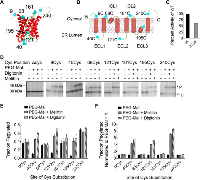
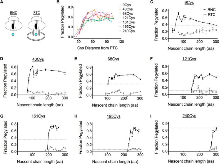
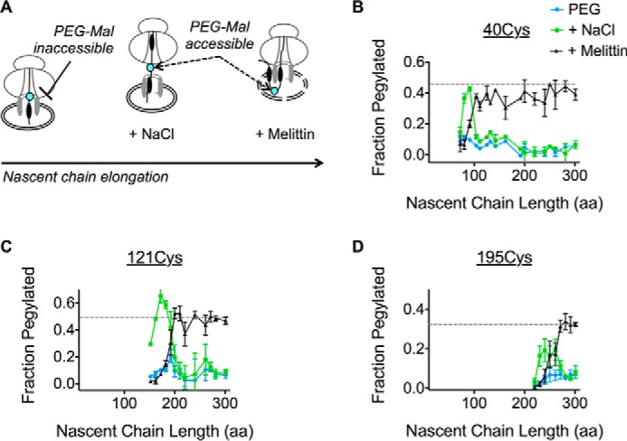
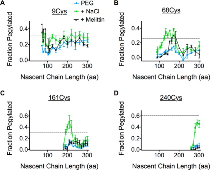
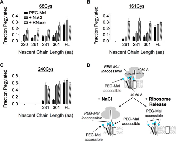
Transmembrane topology of polytopic membrane proteins (PMPs) is established in the endoplasmic reticulum (ER) by the ribosome Sec61-translocon complex (RTC) through iterative cycles of translocation initiation and termination. It remains unknown, however, whether tertiary folding of transmembrane domains begins after the nascent polypeptide integrates into the lipid bilayer or within a proteinaceous environment proximal to translocon components. To address this question, we used cysteine scanning mutagenesis to monitor aqueous accessibility of stalled translation intermediates to determine when, during biogenesis, hydrophilic peptide loops of the aquaporin-4 (AQP4) water channel are delivered to cytosolic and lumenal compartments. Results showed that following ribosome docking on the ER membrane, the nascent polypeptide was shielded from the cytosol as it emerged from the ribosome exit tunnel. Extracellular loops followed a well defined path through the ribosome, the ribosome translocon junction, the Sec61-translocon pore, and into the ER lumen coincident with chain elongation. In contrast, intracellular loops (ICLs) and C-terminalresidues exited the ribosome into a cytosolically shielded environment and remained inaccessible to both cytosolic and lumenal compartments until translation was terminated. Shielding of ICL1 and ICL2, but not the C terminus, became resistant to maneuvers that disrupt electrostatic ribosome interactions. Thus, the early folding landscape of polytopic proteins is shaped by a spatially restricted environment localized within the assembled ribosome translocon complex.

Keywords: aquaporin; endoplasmic reticulum (ER); membrane protein; polytopic membrane protein; protein folding; protein translocation; ribosome function; ribosome translocon complex; sec61 translocon.


Figures

FIGURE 1.
FIGURE 1.
AQP4 topology is unaffected by single cysteine (Cys) substitution and is accurately predicted by PEG-Mal accessibility. A, crystal structure of AQP4 monomer showing sites where single Cys residues (cyan) were incorporated into the AQP4 Δ6Cys mutant (modified from Protein Data Bank code 3gd8) (83). B, two-dimensional schematic showing topology of transmembrane segments and single Cys substitution as in panel A. C, AQP4 Δ6Cys mutant function as measured by osmotic water permeability. D, SDS-PAGE analysis of AQP4 Cys mutants treated with PEG-Mal 5,000. Residues predicted to be cytosolic were modified by PEG-Mal prior to permeabilization. Lumenal residues were pegylated upon addition of digitonin or melittin. Pegylated (P) and unpegylated (U) bands are indicated by single (*) and double asterisks (**), respectively. E, quantified pegylation efficiency from SDS-PAGE in panel D, showing fraction pegylated data calculated from pixel intensity of (P/U + P). F, data from panel E, showing fraction pegylated data normalized to PEG-Mal only.
FIGURE 2.
FIGURE 2.
AQP4 targeting to the ER membrane. A, schematic indicating truncation site relative to position of transmembrane segments in ribosome-attached intermediates. B, SDS-PAGE of AQP4 Δ6Cys truncated integration intermediates showing total translation products (T), supernatant (S), and membrane pellet (P) fractions. Peptidyl-tRNA bands are indicated by the downward arrow (▾). C, quantified fraction of nascent chains in Panel B that remained associated with the microsomes after pelleting ±NaCl, as indicated. In the absence of high salt (open circles), the nascent chain was targeted to the membrane at 72 aa.
FIGURE 3.
FIGURE 3.
The ribosome shields membrane-targeted AQP4. A, schematic showing organization of RNC complexes and membrane-bound RTCs containing the nascent chain with the Cys probe (cyan). RNCs and RTCs were generated from truncated mRNAs in the absence and presence of CRMs, respectively. B, pegylation of each single Cys mutant RNC was plotted as a function of Cys probe distance from the PTC. C-I, cysteines were inaccessible to PEG-Mal in the presence of microsomes. In addition, pegylation of full-length, membrane-targeted AQP4 polypeptide was plotted (dotted line).
FIGURE 4.
FIGURE 4.
Progressive translocation of AQP4 ECLs into the ER lumen. A, schematic showing location of the Cys probe (cyan) within the assembled RTC and effect of high salt (NaCl) on ribosome translocon junction versus melittin on ER membrane integrity. B-D, pegylation of the indicated Cys residues plotted as a function of nascent chain length. For each sample, pegylation was performed in intact microsomes (blue), following the addition of 0.5 m NaCl (green) or melittin (black). Data show that ECL1, ECL2, and ECL3 remain inaccessible to bulk cytosol as they traverse the RTC into the ER lumen.
FIGURE 5.
FIGURE 5.
The ribosome continuously shields multiple ICLs throughout synthesis. A–D, pegylation profiles for Cys residues in the presence of PEG-Mal only (blue), with the addition of high salt (green) or melittin (black). Relative pegylation of the full-length AQP4 with PEG-Mal only is also shown (dotted line).
FIGURE 6.
FIGURE 6.
Salt-insensitive shielding of ICL1 and ICL2 is ribosome dependent. A and B, pegylation of ICL1 (68Cys) and ICL2 (161Cys) at longer truncations following ribosome removal with RNase restores pegylation to a level similar to full-length (FL), membrane-integrated AQP4. C, the C terminus exhibits salt-sensitive pegylation that is not further effected by RNase treatment. D, schematic of salt-insensitive ICL1 and ICL2 shielding and C terminus salt-sensitive shielding by the RTC, which supports a model of TM retention within or near the translocon until translation termination.

References

    1. Skach W. R. (2009) Cellular mechanisms of membrane protein folding. Nat. Struct. Mol. Biol. 16, 606–612 - PMC - PubMed
    1. Alder N. N., and Johnson A. E. (2004) Cotranslational membrane protein biogenesis at the endoplasmic reticulum. J. Biol. Chem. 279, 22787–22790 - PubMed
    1. Hegde R. S., and Lingappa V. R. (1997) Membrane protein biogenesis: regulated complexity at the endoplasmic reticulum. Cell 91, 575–582 - PubMed
    1. White S. H., and von Heijne G. (2004) The machinery of membrane protein assembly. Curr. Opin. Struct. Biol. 14, 397–404 - PubMed
    1. Walter P., and Lingappa V. R. (1986) Mechanism of protein translocation across the endoplasmic reticulum membrane. Annu. Rev. Cell Biol. 2, 499–516 - PubMed

Publication types

MeSH terms

Associated data

LinkOut - more resources