Review
. 2015 Sep;40(9):526-34.
doi: 10.1016/j.tibs.2015.07.002. Epub 2015 Aug 5.

Deciphering voltage-gated Na(+) and Ca(2+) channels by studying prokaryotic ancestors

Affiliations
Review

Deciphering voltage-gated Na(+) and Ca(2+) channels by studying prokaryotic ancestors

William A Catterall et al. Trends Biochem Sci. 2015 Sep.

Abstract

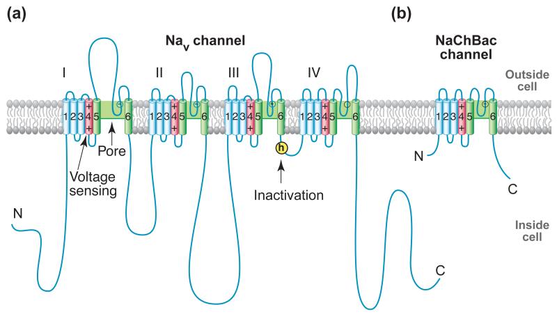
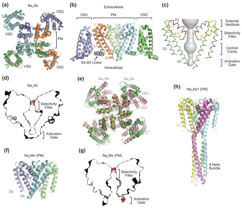
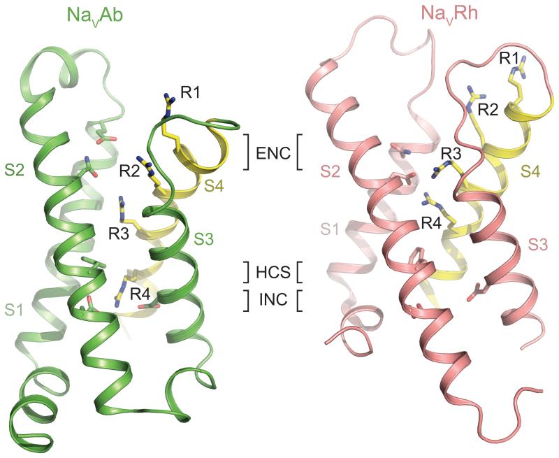
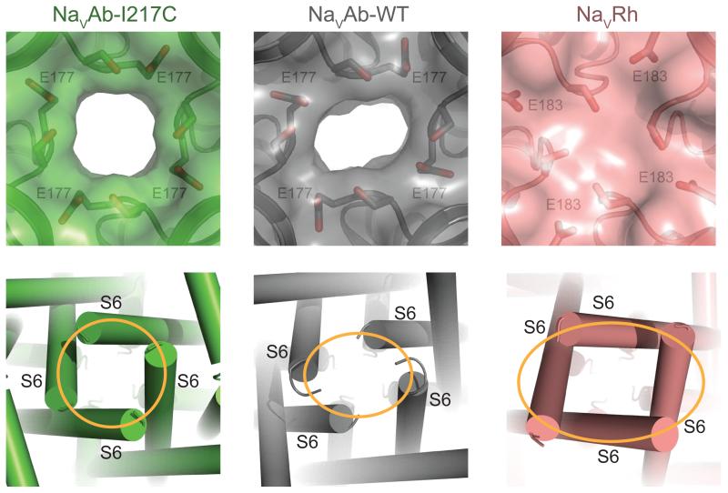
Voltage-gated sodium channels (NaVs) and calcium channels (CaVs) are involved in electrical signaling, contraction, secretion, synaptic transmission, and other physiological processes activated in response to depolarization. Despite their physiological importance, the structures of these closely related proteins have remained elusive because of their size and complexity. Bacterial NaVs have structures analogous to a single domain of eukaryotic NaVs and CaVs and are their likely evolutionary ancestor. Here we review recent work that has led to new understanding of NaVs and CaVs through high-resolution structural studies of their prokaryotic ancestors. New insights into their voltage-dependent activation and inactivation, ion conductance, and ion selectivity provide realistic structural models for the function of these complex membrane proteins at the atomic level.

Keywords: Na(V)Ab; NaChBac; selectivity filter; slow inactivation; voltage sensor; voltage-gated calcium channel; voltage-gated sodium channel.


Figures

Figure 1
Figure 1. NaV Channel Structure
(a) Two-dimensional schematic map of NaV channel structure and function. The α subunit of NaV1.2 channels is illustrated as a transmembrane folding diagram in which cylinders represent transmembrane alpha helices and lines represent connecting amino acid sequences in proportion to their length. The roman numerals indicate the four homologous domains and the Arabic numerals are used to label the six transmembrane helices. The S4 helices are colored in red with “+” signs indicating gating charges. The S5-S5 helices are colored in green and the small white circles indicate key residues in the selectivity filter with “+” and “−“ signs indicating their charge states. The yellow circle with an “h” indicates inactivation gate. (b) Schematic map of the bacterial NaChBac channel, which contains the minimal functional elements of a single homologous domain in a mammalian NaV channel.
Figure 2
Figure 2. Overall Structures of Prokaryotic NaV Channels
(a) Structure of the NaVAb bacterial NaV channel determined at 2.7Å resolution and viewed from the extracellular side [9]. The four subunits of the homotetrameric channel are shown in different colors. The central pore is surrounded by four VSMs. (b) Side view of NaVAb with the same coloring scheme as shown in (a). S5 and S6 helices of one subunit (slate) and the VSM of another subunit (cyan) are omitted for clarity. (c) Architecture of the NaVAb pore with pore volume shown in grey and the high-strength-field (HSF) site residue Glu177 shown in sticks. P and P2 indicate the P and P2 helices. (d) Cross-section of the NaVAb pore showing the closed activation gate. The selectivity filter is shown with electrostatic surface potential colored from −50 to 50 kT (red to blue). (e) Superposition of NaVRh and NaVAb with their PMs superimposed [9, 11]. The VSM of the two channels packs against the PM at different angles. (f) Structure of the NaVMs pore module [13]. (g) Cross-section of the NaVMs pore showing the open conformation of its activation gate [18]. The selectivity filter is shown with electrostatic surface potential colored from −50 to 50 kT (red to blue). (h) Structure of the NaVAe1 pore with well-resolved C-terminal domain forming a four-helix bundle [15].
Figure 3
Figure 3. A Comparison of the Voltage-sensor Domains of NaVAb and NaVRh
The VSMs of NaVAb (left, green) and NaVRh (right, salmon) are shown in ribbon diagram [9, 11]. The sliding S4 helix is highlighted in yellow with the side chains of the four gating charge arginine residues (R1-R4) shown in sticks. Key residues forming the extracellular negative cluster (ENC), intracellular negative cluster (INC), and hydrophobic constriction site (HCS) are shown in sticks. The R4 residue of NaVAb and NaVRh interacts with the INC and ENC residues, respectively.
Figure 4
Figure 4. Structural Basis of Slow Inactivation
Specific structural features of NaVAb-WT and NaVRh, including a distorted selectivity filter and the breakdown of the four-fold symmetry of the pore, as expected for the slow inactivation state. Top panel: Close-up views of the extracellular entrance of NaVAb-I217C (green, symmetric), NaVAb-WT (grey, asymmetric), and NaVRh (salmon, collapsed) with semi-transparent surface representation of the three channels [9-11]. The high-strength-field site glutamate residue in each structure along with its nearby serine residue is shown in sticks. Bottom panel: Close-up view of the intracellular closed activation gate of NaVAb-I217C (symmetric, orange circle), NaVAb-WT (asymmetric, orange oval), and NaVRh (asymmetric, orange oval). .
Figure 5
Figure 5. Selectivity Filter of NaVAb
(a) Close-up view of the extracellular entrance of NaVAb-I217C with the high-strength-field site, Glu177, highlighted in yellow [9]. (b) Side view of the NaVAb-I217C selectivity filter showing Glu177 (yellow) and the backbone carbonyls of Leu176 and Thr175, all of which are involved in selecting and permeating partially hydrated sodium ions. (c) Superposition of NaVAb and a K+-channel (PDB code 1K4C) selectivity filter. The structural alignment, which is based on the common P-helices of the two channels, highlights the significant differences in the width of the two selectivity filters. (d) Different conformational states of the four high-strength-field site Glu177 residues captured in equilibrium molecular dynamics simulation of NaVAb in a hydrated lipid bilayer with Na+ moving in and out of the pore [40]. The side chains of Glu177 point either out toward the mouth of the selectivity filter or into the lumen (0-4 indicate the number of Glu side chains dunked; Na+ ions not shown). (e) Axial distribution of Na+ atoms in the selectivity filter and central cavity of NaVAb. Three distinguishable states are highlighted in which Na+ is directly bound to Glu177 (green), to both Glu177 and the backbone carbonyl of Leu176 (yellow), or to neither (brown). (d) and (e) are adapted from reference [40].
Figure 6
Figure 6. Ca2+ Selectivity and Permeation by CaVAb
(a) Side view of the superimposed selectivity filters of CaVAb and NaVAb [9, 46]. The side chains of the three residues of NaVAb mutated to generate CaVAb are colored in yellow in the CaVAb structure. The backbone structure of the selectivity filters of the two channels are nearly identical. (b) Side view of the CaVAb selectivity filter with three Ca2+ (green spheres) binding sites and their coordinating oxygen atoms. The distances between Ca2+ and the oxygen atoms indicated with dash lines range from 4.0 Å to 5.0 Å. (c) A proposed mechanism of Ca2+ permeation by CaVAb based on a combination of the “knock-out” and “stepwise permeation” models. The selectivity filter oscillates between two proposed ionic occupancy states where Ca2+ ions either bind to position 2, the high-affinity binding site, or position 1 and 3, which bind the ion at lower affinities.

References

    1. Hille B. Ionic Channels of Excitable Membranes. 3rd Ed Sinauer Associates Inc.; Sunderland, MA: 2001.
    1. Tsien RW. Calcium channels in excitable cell membranes. Annu Rev Physiol. 1983;45:341–358. - PubMed
    1. Catterall WA. Voltage-gated calcium channels. Cold Spring Harb Perspect Biol. 2011;3:a003947. - PMC - PubMed
    1. Catterall WA. The molecular basis of neuronal excitability. Science. 1984;223:653–661. - PubMed
    1. Catterall WA. From ionic currents to molecular mechanisms: The structure and function of voltage-gated sodium channels. Neuron. 2000;26:13–25. - PubMed

Publication types

MeSH terms

Substances

LinkOut - more resources