. 2015 Nov 1;407(1):90-102.
doi: 10.1016/j.ydbio.2015.08.004. Epub 2015 Aug 5.

Mammalian target of rapamycin complex 1 (mTORC1) Is required for mouse spermatogonial differentiation in vivo

Affiliations

Mammalian target of rapamycin complex 1 (mTORC1) Is required for mouse spermatogonial differentiation in vivo

Jonathan T Busada et al. Dev Biol. .

Abstract

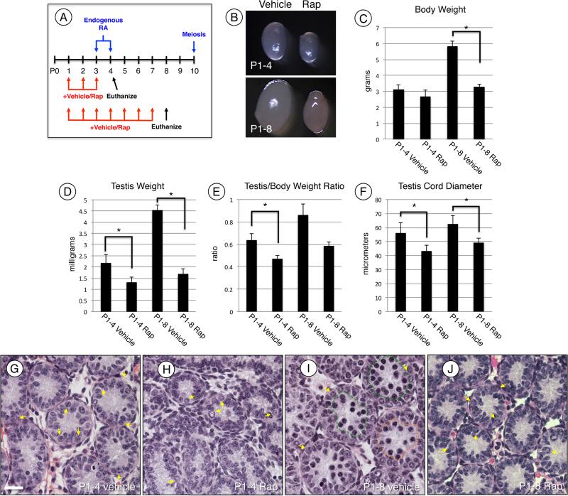
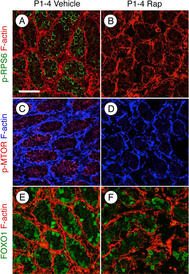
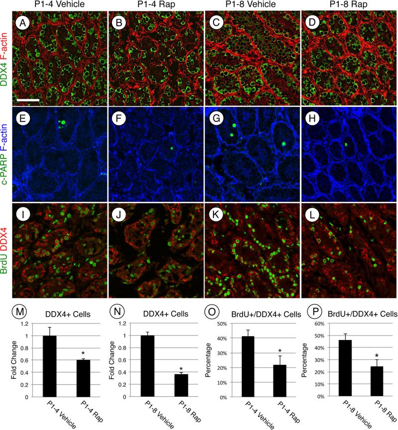
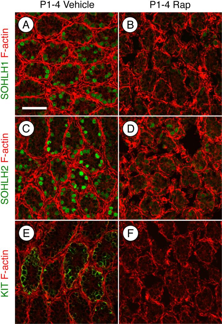
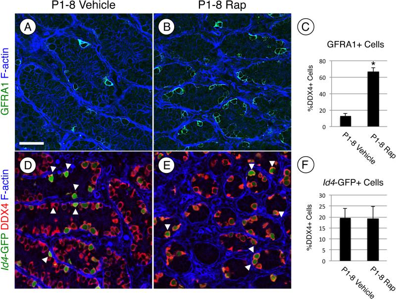
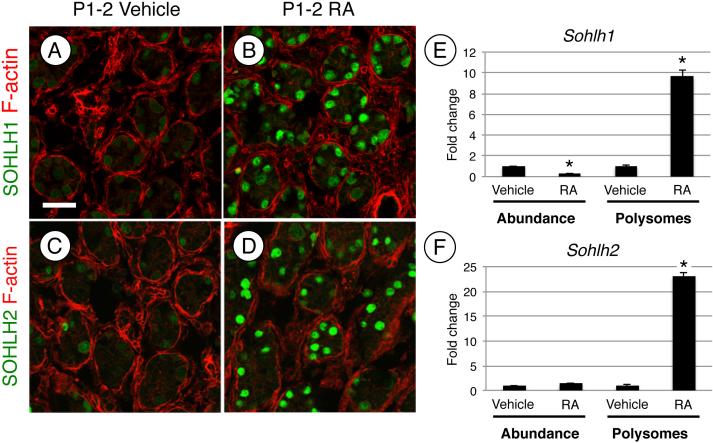
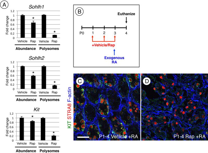
Spermatogonial stem cells (SSCs) must balance self-renewal with production of transit-amplifying progenitors that differentiate in response to retinoic acid (RA) before entering meiosis. This self-renewal vs. differentiation spermatogonial fate decision is critical for maintaining tissue homeostasis, as imbalances cause spermatogenesis defects that can lead to human testicular cancer or infertility. A great deal of effort has been exerted to understand how the SSC population is maintained. In contrast, little is known about the essential program of differentiation initiated by retinoic acid (RA) that precedes meiosis, and the pathways and proteins involved are poorly defined. We recently reported a novel role for RA in stimulating the PI3/AKT/mTOR kinase signaling pathway to activate translation of repressed mRNAs such as Kit. Here, we examined the requirement for mTOR complex 1 (mTORC1) in mediating the RA signal to direct spermatogonial differentiation in the neonatal testis. We found that in vivo inhibition of mTORC1 by rapamycin blocked spermatogonial differentiation, which led to an accumulation of undifferentiated spermatogonia. In addition, rapamycin also blocked the RA-induced translational activation of mRNAs encoding KIT, SOHLH1, and SOHLH2 without affecting expression of STRA8. These findings highlight dual roles for RA in germ cell development - transcriptional activation of genes, and kinase signaling to stimulate translation of repressed messages required for spermatogonial differentiation.

Keywords: Retinoic acid; Spermatogenesis; Spermatogonia; Testis; Translation; mTOR.


Figures

Figure 1
Figure 1. MTORC1 inhibition blocks spermatogonial differentiation
(A) Experimental design for treating mice with rapamycin in vivo. Mice were treated with vehicle or rapamycin once daily starting at P1 and euthanized 24h after last treatment at P4 or P8. (B) Representative images of testes from vehicle (left) and rapamycin (right) treated mice euthanized at P4 (top) and P8 (bottom). (C-E) Mice treated with vehicle or rapamycin were euthanized at P4 or P8. Prior to euthanasia total body weights were collected (C), following euthanasia total testis weights were collected (D). Testis weights were normalized to body weights and expressed as a ratio (E). (F) Quantitation of testis cord diameter of mice treated with vehicle or rapamycin and euthanized at P4 or P8. (G-J) H&E staining of mice treated with vehicle (G and I) or rapamycin (H and J) and euthanized at P4 (G and H) or P8 (I and J). Yellow arrows indicate spermatogonia, and green and orange lines encircle preleptotene and leptotene spermatocytes, respectively (I). Scale bar = 40 μM. Asterisks indicate statistical significance with P<0.01.
Figure 2
Figure 2. Treatment with rapamycin inhibits mTORC1 activity without affecting AKT
(A-F) Immunostaining of testis sections from mice treated with vehicle (A, C, and E) or rapamycin (B, D, and F) and euthanized at P4. Sections were stained with anti- phosphorylated RPS6 (A and B), anti- phosphorylated EIF4EBP1 (C and D), or total FOXO1 (E and F). F-actin was stained with phalloidin (red) to visualize testis cords. Scale bar = 50 μM.
Figure 3
Figure 3. MTORC1 is required for postnatal expansion of the germ cell population
(A-L) Immunostaining of testis sections from mice treated with vehicle (A, C, E, G, I, and K) or rapamycin (B, D, F, H, J, and L) and euthanized at P4 (A, B, E, F, I, and J) or P8 (C, D, G, H, K, and L). Sections were stained with anti-DDX4 (A-D), anti-cleaved PARP1 (E-H), or double labeled with anti-BrdU (green, I-L) and anti-DDX4 (red, I-L). F-actin was stained with phalloidin (red, A-D or blue, E-H). Quantitation of the number of DDX4+(M and N) or the number of BrdU+/DDX4+ (O and P) cells from testes treated starting at P1 with vehicle or rapamycin and then euthanized at P4 (M and O) or at P8 (N and P). Asterisks indicate statistical significance with P≤0.01. Scale bar = 50 μM.
Figure 4
Figure 4. MTOR activation is required for induction of SOHLH1, SOHLH2, and KIT protein
Immunostaining of mice treated with vehicle (A, C, and E) or rapamycin (B, D, and F) and euthanized at P4. Sections were stained with anti-SOHLH1 (A-B), anti-SOHLH2 (C-D), or anti-KIT (E-F). Phalloidin (red) was added to visualize testis cords. Scale bar = 50 μM.
Figure 5
Figure 5. Inhibiting mTORC1 activation increases the number of undifferentiated spermatogonia
(A, B, D, and E) Immunostaining was performed on testis sections from mice treated with vehicle (A and D) or rapamycin (B and E) and euthanized at P8. (A-C) Testis sections from CD-1 mice were stained with anti-GFRA1 (green, A and B) and F-actin was stained with phalloidin (blue) to visualize testis cords. The number of GFRA1+ germ cells in vehicle- and rapamycin-treated testes were quantitated and reported as a fold change (C). (D-F) Transgenic Id4-GFP mice were treated with vehicle or rapamycin, and immunostaining was performed on testes. Green represents GFP epifluoresence, and sections were labeled with anti-DDX4 (red). White arrows indicate GFP bright spermatogonia (D and E). The number of GFP bright cells were quantitated and represented as a percentage of the DDX4+ cells (C). Scale bar = 40 μM. Asterisks indicate statistical significance with P<0.01.
Figure 6
Figure 6. RA induces expression of SOHLH1 and SOHLH2 protein
(A-D) Immunostaining of testis sections from mice treated at P1 with vehicle (A and C) or RA (B and D) and euthanized 24h later (at P2). Sections were stained with anti-SOHLH1 (A and B) or anti-SOHLH2 (C and D), and F-actin was stained with phalloidin (red) to visualize testis cords. QRT-PCR was performed on RNA isolated from whole testis lysate of mice treated with vehicle or RA, and total Sohlh1 and Sohlh2 mRNA levels were measured (left side, E and F). Messenger RNAs were separated by ribosome occupancy, fractions containing heavy polysomes were pooled, and qRT-PCR was performed to quantify polysome-associated Sohlh1 and Sohlh2 (right side, E and F). Scale bar = 30 μM. Asterisks indicate statistical significance with P<0.01.
Figure 7
Figure 7. RA signaling through mTORC1 is required for induction of KIT but not STRA8
(A) Message levels for Sohlh1, Sohlh2, and Kit were measured by qRT-PCR using whole testis RNA of mice treated with vehicle or rapamycin and then euthanized at P4. Sohlh1, Sohlh2, and Kit mRNAs associated with polysomes were pooled, isolated, and quantitated by qRT-PCR. (B) Mice were treated daily starting at P1 with vehicle or rapamycin. At P3, mice were given a single exogenous injection of RA and euthanized 24h later at P4. (C and D) Immunostaining of testis sections of mice treated with vehicle (C) or rapamycin (D) and RA and then euthanized at P4. Sections were labeled with KIT (green) and STRA8 (red), F-actin was stained with phalloidin to visualize testis cords (in blue). Scale bar = 40 μM. Asterisks indicate statistical significance with P<0.01.
Figure 8
Figure 8. RA signaling through PI3K/AKT/mTOR is required for spermatogonia differentiation
Specific mRNAs are inefficiently translated (repressed) in undifferentiated germ cells. RA signaling through a kinase (non-genomic) signaling pathway activates the PI3K/AKT/mTORC1 signaling network to induce efficient translation of genes (e.g. Kit, Sohlh1, and Sohlh2) that are required for differentiation. Rapamycin inhibition of mTORC1 prevents RA induced efficient translation, and blocks spermatogonia differentiation.

References

    1. Bowles J, Knight D, Smith C, Wilhelm D, Richman J, Mamiya S, Yashiro K, Chawengsaksophak K, Wilson MJ, Rossant J, Hamada H, Koopman P. Retinoid signaling determines germ cell fate in mice. Science. 2006;312:596–600. - PubMed
    1. Koubova J, Menke DB, Zhou Q, Capel B, Griswold MD, Page DC. Retinoic acid regulates sex-specific timing of meiotic initiation in mice. Proc Natl Acad Sci U S A. 2006;103:2474–2479. - PMC - PubMed
    1. Busada JT, Kaye EP, Renegar RH, Geyer CB. Retinoic acid induces multiple hallmarks of the prospermatogonia-to-spermatogonia transition in the neonatal mouse. Biol Reprod. 2014;90:64. - PMC - PubMed
    1. Niedenberger BA, Busada JT, Geyer CB. Marker expression reveals heterogeneity of spermatogonia in the neonatal mouse testis. Reproduction. 2015;149:329–338. - PMC - PubMed
    1. Snyder EM, Small C, Griswold MD. Retinoic acid availability drives the asynchronous initiation of spermatogonial differentiation in the mouse. Biol Reprod. 2010;83:783–790. - PMC - PubMed

Publication types

MeSH terms

Substances