. 2015 Dec;76(2):244-252.
doi: 10.1016/j.cyto.2015.07.027. Epub 2015 Aug 5.

Interleukin-6 inhibits apoptosis of exocrine gland tissues under inflammatory conditions

Affiliations

Interleukin-6 inhibits apoptosis of exocrine gland tissues under inflammatory conditions

Jing Zhou et al. Cytokine. 2015 Dec.

Abstract

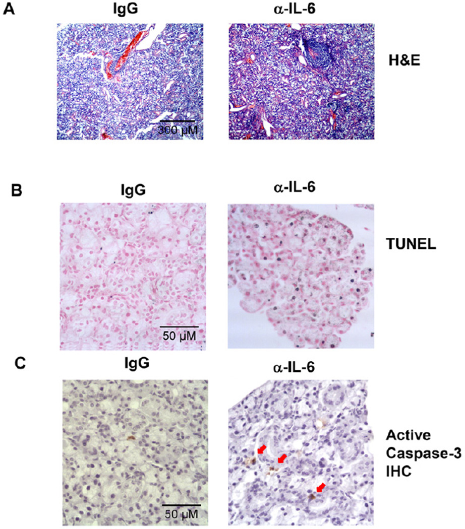
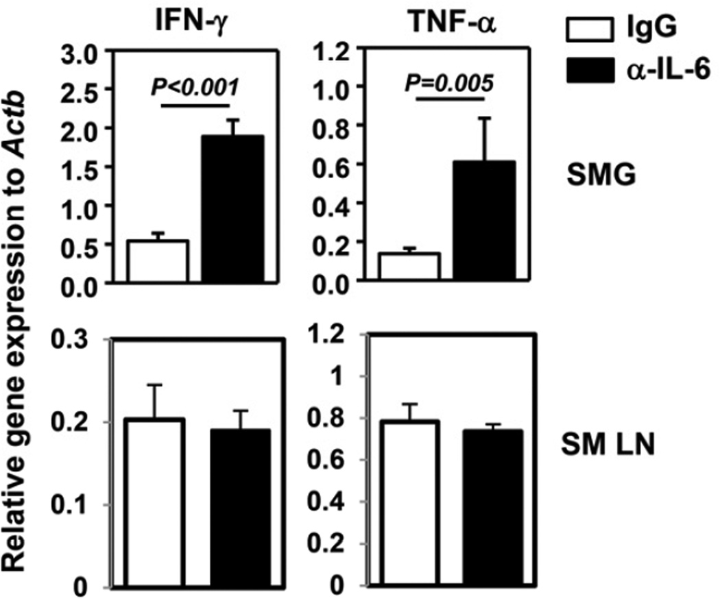
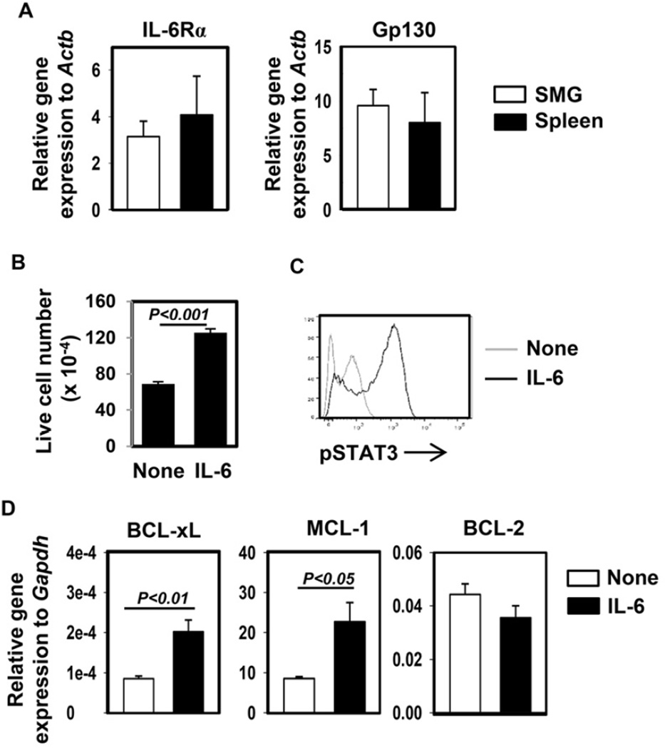
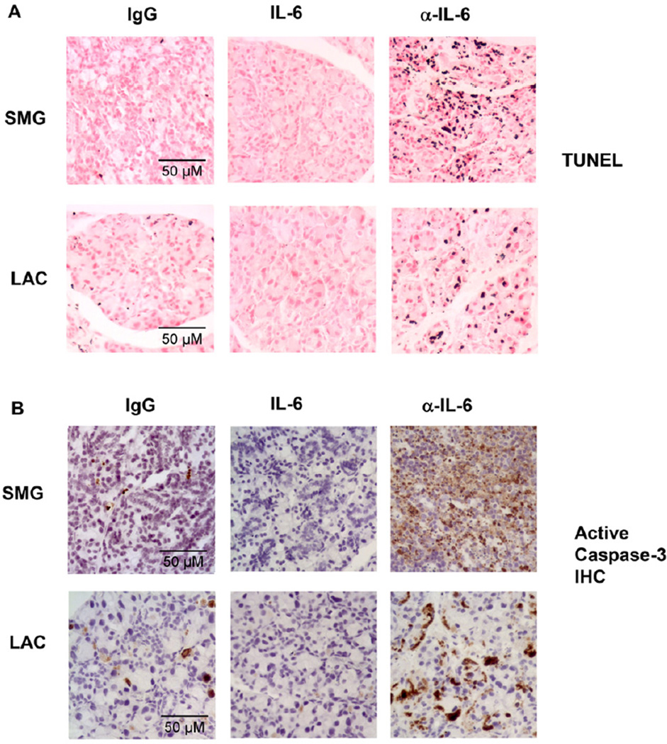
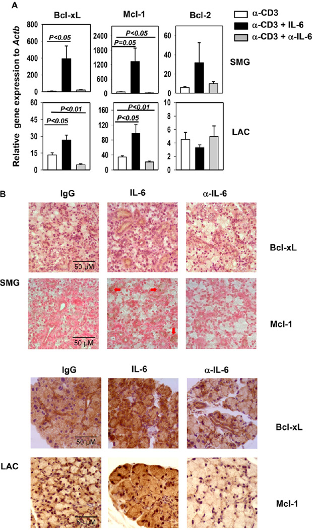
Interleukin (IL)-6 is a multi-functional cytokine that can either promote or suppress tissue inflammation depending on the specific disease context. IL-6 is elevated in the exocrine glands and serum of patients with Sjögren's syndrome (SS), but the specific role of IL-6 in the pathogenesis of this disease has not been defined. In this study, we showed that IL-6 expression levels were increased with age in C56BL/6.NOD-Aec1Aec2 mice, a primary SS model, and higher than the control C57BL/6 mice. To assess the role of IL-6 during the immunological phase of SS development, a neutralizing anti-IL-6 antibody was administered into 16 week-old female C56BL/6.NOD-Aec1Aec2 mice, 3 times weekly for a consecutive 8 weeks. Neutralization of endogenous IL-6 throughout the immunological phase of SS development led to increased apoptosis, caspase-3 activation, leukocytic infiltration, and IFN-γ- and TNF-α production in the salivary gland. To further determine the effect of IL-6 on the apoptosis of exocrine gland cells, recombinant human IL-6 or the neutralizing anti-IL-6 antibody was injected into female C57BL/6 mice that received concurrent injection of anti-CD3 antibody to induce the apoptosis of exocrine gland tissues. Neutralization of IL-6 enhanced, whereas administration of IL-6 inhibited apoptosis and caspase-3 activation in salivary and lacrimal glands in this model. The apoptosis-suppressing effect of IL-6 was associated with up-regulation of Bcl-xL and Mcl-1 in both glands. Moreover, IL-6 treatment induced activation of STAT3 and up-regulated Bcl-xL and Mcl-1 gene expression in a human salivary gland epithelial cell line. In conclusion, IL-6 inhibits the apoptosis of exocrine gland tissues and exerts a tissue-protective effect under inflammatory conditions including SS. These findings suggest the possibility of using this property of IL-6 to preserve exocrine gland tissue integrity and function under autoimmune and inflammatory conditions.

Keywords: Apoptosis; Exocrine gland; Inflammation; Interleukin-6; Sjögren’s syndrome.


Figures

Figure 1
Figure 1. Expression of IL-6 and its receptor in the SMG of B6.NOD-Aec mice
(A) IL-6, IL-6Rα and gp130 mRNA levels in the SMG of B6 or B6.NOD-Aec mice aged 10–12, 16–18 and 24 weeks. Expression levels were presented relative to that of β-actin. (B) Serum IL-6 concentrations in the same mice described above. Data are the average of analyses of at least 6 mice for each group from 3 independent experiments. The histograms and error bars represent the average and SEM, respectively.
Figure 2
Figure 2. Neutralization of IL-6 in B6.NOD-Aec mice leads to increased inflammation and apoptosis in SMG
Anti-IL-6 or control IgG was injected to 16 week-old B6.NOD-Aec mice, 3 times weekly for 8 weeks. (A) H&E staining of SMG sections showing leukocytic foci. Original magnification: ×10. (B) In situ TUNEL staining of apoptotic cells in SMG sections. Original magnification: ×40. (C) Immunohistochemical staining of active caspase-3 in SMG sections. Data are representative of the analyses of 9 mice for each group from 5 independent experiments. Original magnification: ×40.
Figure 3
Figure 3. Neutralization of IL-6 in B6.NOD-Aec mice leads to increased IFN-γ and TNF-α expression in the SMG
IFN-γ and TNF-α mRNA levels in the submandibular gland (SMG) and submandibular draining lymph nodes (SM LN) of B6.NOD-Aec mice injected with anti-IL-6 or control IgG as described in Figure 2. Expression levels were presented relative to that of β-actin. Data are the average of the analyses of 7 mice for each group from 4 independent experiments. The histograms and error bars represent the average and SEM, respectively.
Figure 4
Figure 4. IL-6 activates STAT3 and up-regulates expression of anti-apoptotic molecules in HSG cells
(A) Numbers of viable HSG cells after being cultured in vitro in the presence or absence of rh IL-6 for 5 days. Data are the average of 6 independent samples from 2 experiments. The histograms and error bars represent the average and SEM, respectively. (B) Flow cytometry of intracellular p-STAT3 in HSG cells transiently stimulated with rh IL-6. Data are representative of 4 independent analyses from 4 experiments. (C) Bcl-xL, Bcl-2 and Mcl-1 mRNA levels in HSG cells cultured with or without rh IL-6 for 1 day, presented relative to that of human Gapdh gene. Data are the average the analyses of 4 independent samples for each group. The histograms and error bars represent the average and SEM, respectively.
Figure 5
Figure 5. IL-6 inhibits exocrine gland cell death induced by in vivo anti-CD3 administration
B6 mice were injected with anti-CD3 in the presence or absence of rh IL-6 or anti-IL-6. (A) In situ TUNEL staining of apoptotic cells in SMG and LAC sections. (B) Immunohistochemical staining of active caspase-3 in SMG and LAC sections. Data are representative of the analyses of 8 mice for each group from 3 independent experiments. Original magnification: × 40.
Figure 6
Figure 6. IL-6 up-regulates Bcl-xL and Mcl-1 expression in exocrine glands in anti-CD3-treated mice
(A) Bcl-xL, Mcl-1 and Bcl-2 mRNA levels in the SMG and LAC of B6 mice treated with anti-CD3 with or without rh IL-6 or anti-IL-6 as described in Figure 5. Expression levels were presented relative to that of β-actin. The histograms and error bars represent the average and SEM, respectively. (B) Immunohistochemical staining of Bcl-xL and Mcl-1 in SMG and LAC sections. Data are the average of or representative of the analyses of 7–8 mice each group from 3 independent experiments. Original magnification: × 40.

References

    1. Neurath MF, Finotto S. IL-6 signaling in autoimmunity, chronic inflammation and inflammation-associated cancer. Cytokine Growth Factor Rev. 22:83–89. - PubMed
    1. Danese S, Gao B. Interleukin-6: a therapeutic Jekyll and Hyde in gastrointestinal and hepatic diseases. Gut. 2010;59:149–151. - PMC - PubMed
    1. Gabay C. Interleukin-6 and chronic inflammation. Arthritis research & therapy. 2006;8(Suppl 2):S3. - PMC - PubMed
    1. Fonseca JE, Santos MJ, Canhao H, Choy E. Interleukin-6 as a key player in systemic inflammation and joint destruction. Autoimmun Rev. 2009;8:538–542. - PubMed
    1. Lindroos J, Svensson L, Norsgaard H, Lovato P, Moller K, Hagedorn PH, et al. IL-23-mediated epidermal hyperplasia is dependent on IL-6. The Journal of investigative dermatology. 131:1110–1118. - PubMed

Publication types

MeSH terms