

Case Reports
doi: 10.1292/jvms.15-0194.
Epub 2015 Aug 8.
Computed tomographic images of discospondylitis in a calf
Affiliations
- PMID: 26256229
- PMCID: PMC4710732
- DOI: 10.1292/jvms.15-0194
Item in Clipboard
Case Reports
Computed tomographic images of discospondylitis in a calf
J Vet Med Sci.
2016 Jan.
Abstract
A 2-month-old male Japanese Black calf was presented with a 30-day history of progressive ataxia. Antemortem examination using computed tomography (CT) revealed narrowing of the disc spaces due to destruction of intervertebral structures between the first and second thoracic vertebrae and between the second and third thoracic vertebrae. Osteolysis was evident as irregular hypoattenuating lesions within the opposing end plates of the first, second and third thoracic vertebrae. Pseudomonas aeruginosa was detected as the causative bacteria, and discospondylitis was diagnosed. To the best of our knowledge, this is the first bovine case report describing the application of CT for the diagnosis of discospondylitis.
Figures

Lateral radiograph of the cervical-thoracic vertebrae. Osteolysis is evident in the
caudal part of the end plate of the first thoracic vertebra. Osteolytic changes cause
reduced length of the second thoracic vertebra. Bar=25 mm.

Transverse computed tomography of the thoracic vertebra. (A) An oval-shaped focal
lesion is evident at the center of the caudal part of the end plate of the first
thoracic vertebra. (B) The hypoattenuating lesion has destroyed the ventral edge of the
cranial part of the end plate of the second thoracic vertebra. No bony proliferation is
apparent in the first or second thoracic vertebrae. Bar=10 mm.
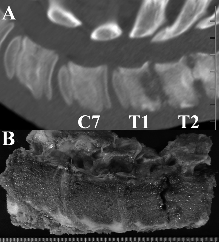
Sagittal reconstructed computed tomography (A) and gross appearance (B) of the
cervical-thoracic vertebrae. A deeply concave osteolytic lesion, showing hypoattenuation
on CT, is filled with dark-red necrotic material within the caudal part of the end plate
of the first thoracic vertebra. Osteolysis is grossly evident within the cranial and
caudal parts of the end plate of the second thoracic vertebra, which appears as a
shortened vertebral body with irregular edges at either end on CT. Bar=10 mm.

Histopathology of the cranial end plate of the second thoracic vertebral bone. The bony
architecture is replaced by severe inflammatory cells. These cells mainly consist of
neutrophils and spindle-shaped fibroblastic cells (inset). The remaining cartilaginous
tissue of the vertebra is located at the lower right. Bar=200 µm.
References
-
- Betbeze C., McLaughlin R.2002. Canine diskospondylitis: Its etiology, diagnosis, and treatment. Vet. Med. 97: 673–681.
Publication types
MeSH terms
LinkOut - more resources
Full Text Sources
Other Literature Sources
Medical