

Studying polyglutamine diseases in Drosophila
- PMID: 26257024
- PMCID: PMC4644473
- DOI: 10.1016/j.expneurol.2015.08.002
Studying polyglutamine diseases in Drosophila
Abstract
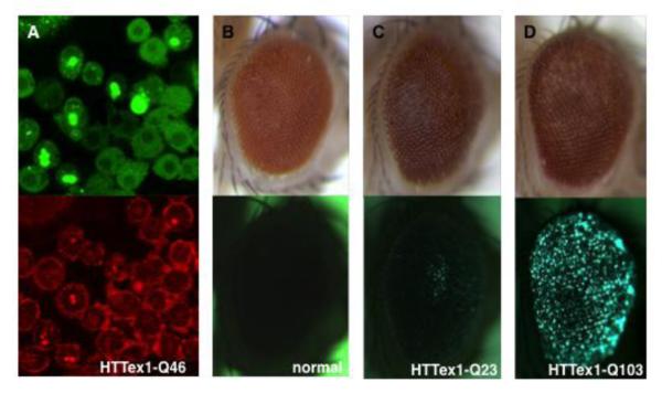
Polyglutamine (polyQ) diseases are a family of dominantly transmitted neurodegenerative disorders caused by an abnormal expansion of CAG trinucleotide repeats in the protein-coding regions of the respective disease-causing genes. Despite their simple genetic basis, the etiology of these diseases is far from clear. Over the past two decades, Drosophila has proven to be successful in modeling this family of neurodegenerative disorders, including the faithful recapitulation of pathological features such as polyQ length-dependent formation of protein aggregates and progressive neuronal degeneration. Additionally, it has been valuable in probing the pathogenic mechanisms, in identifying and evaluating disease modifiers, and in helping elucidate the normal functions of disease-causing genes. Knowledge learned from this simple invertebrate organism has had a large impact on our understanding of these devastating brain diseases.
Keywords: Atrophin-1; DRPLA; Dentatorubral-pallidoluysian atrophy; Drosophila model; HD; HTT; Huntingtin; Huntington's disease; Machado–Joseph disease; PolyQ diseases; Polyglutamine diseases; SBMA; SCA1; Spinobulbar muscular atrophy; Spinocerebellar ataxia.
Copyright © 2015 Elsevier Inc. All rights reserved.
Figures
Similar articles
-
Polyglutamine (PolyQ) diseases: genetics to treatments.Cell Transplant. 2014;23(4-5):441-58. doi: 10.3727/096368914X678454. Cell Transplant. 2014. PMID: 24816443 Review.
-
[Molecular biology of polyglutamine diseases].Postepy Hig Med Dosw. 2002;56(6):779-88. Postepy Hig Med Dosw. 2002. PMID: 12661407 Review. Polish.
-
Increased aggregation of polyleucine compared with that of polyglutamine in dentatorubral-pallidoluysian atrophy protein.Neurosci Lett. 2013 Sep 27;552:156-61. doi: 10.1016/j.neulet.2013.07.043. Epub 2013 Aug 7. Neurosci Lett. 2013. PMID: 23933208
-
Pathogenesis of SCA3 and implications for other polyglutamine diseases.Neurobiol Dis. 2020 Feb;134:104635. doi: 10.1016/j.nbd.2019.104635. Epub 2019 Oct 24. Neurobiol Dis. 2020. PMID: 31669734 Free PMC article. Review.
-
Autophagy and Polyglutamine Disease.Adv Exp Med Biol. 2020;1207:149-161. doi: 10.1007/978-981-15-4272-5_9. Adv Exp Med Biol. 2020. PMID: 32671744 Review.
Cited by
-
Single cell RNA sequencing of the adult Drosophila eye reveals distinct clusters and novel marker genes for all major cell types.Commun Biol. 2022 Dec 14;5(1):1370. doi: 10.1038/s42003-022-04337-1. Commun Biol. 2022. PMID: 36517671 Free PMC article.
-
Development of a Polymeric Pharmacological Nanocarrier System as a Potential Therapy for Spinocerebellar Ataxia Type 7.Cells. 2023 Nov 30;12(23):2735. doi: 10.3390/cells12232735. Cells. 2023. PMID: 38067163 Free PMC article.
-
A Short History and Description of Drosophila melanogaster Classical Genetics: Chromosome Aberrations, Forward Genetic Screens, and the Nature of Mutations.Genetics. 2017 Jun;206(2):665-689. doi: 10.1534/genetics.117.199950. Genetics. 2017. PMID: 28592503 Free PMC article.
-
The deubiquitinase USP45 inhibits autophagy through actin regulation by Coronin 1B.J Cell Biol. 2025 May 5;224(5):e202407014. doi: 10.1083/jcb.202407014. Epub 2025 Mar 11. J Cell Biol. 2025. PMID: 40067150 Free PMC article.
-
Using Drosophila Motor Mutants to Teach Neurodevelopment in an Undergraduate Neurobiology Lab.J Undergrad Neurosci Educ. 2020 Jun 28;18(2):A93-A101. eCollection 2020 Spring. J Undergrad Neurosci Educ. 2020. PMID: 32848517 Free PMC article.
References
-
- A novel gene containing a trinucleotide repeat that is expanded and unstable on Huntington’s disease chromosomes. The Huntington’s Disease Collaborative Research Group. Cell. 1993;72:971–983. - PubMed
-
- David G, et al. Cloning of the SCA7 gene reveals a highly unstable CAG repeat expansion. Nat Genet. 1997;17:65–70. - PubMed
-
- Imbert G, et al. Cloning of the gene for spinocerebellar ataxia 2 reveals a locus with high sensitivity to expanded CAG/glutamine repeats. Nat Genet. 1996;14:285–291. - PubMed
-
- Kawaguchi Y, et al. CAG expansions in a novel gene for Machado-Joseph disease at chromosome 14q32.1. Nat Genet. 1994;8:221–228. - PubMed
-
- Koob MD, et al. Rapid cloning of expanded trinucleotide repeat sequences from genomic DNA. Nat Genet. 1998;18:72–75. - PubMed
Publication types
MeSH terms
Substances
Grants and funding
LinkOut - more resources
Full Text Sources
Other Literature Sources
Medical
Molecular Biology Databases
Miscellaneous
