. 2015 Aug 18;12(7):1169-83.
doi: 10.1016/j.celrep.2015.07.023. Epub 2015 Aug 6.

Exome Sequence Analysis Suggests that Genetic Burden Contributes to Phenotypic Variability and Complex Neuropathy

Affiliations

Exome Sequence Analysis Suggests that Genetic Burden Contributes to Phenotypic Variability and Complex Neuropathy

Claudia Gonzaga-Jauregui et al. Cell Rep. .

Abstract

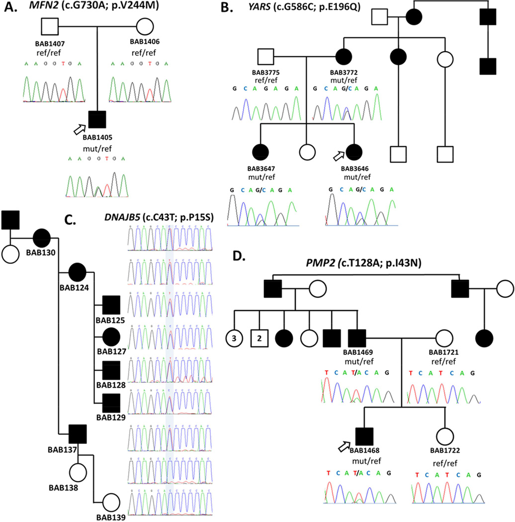
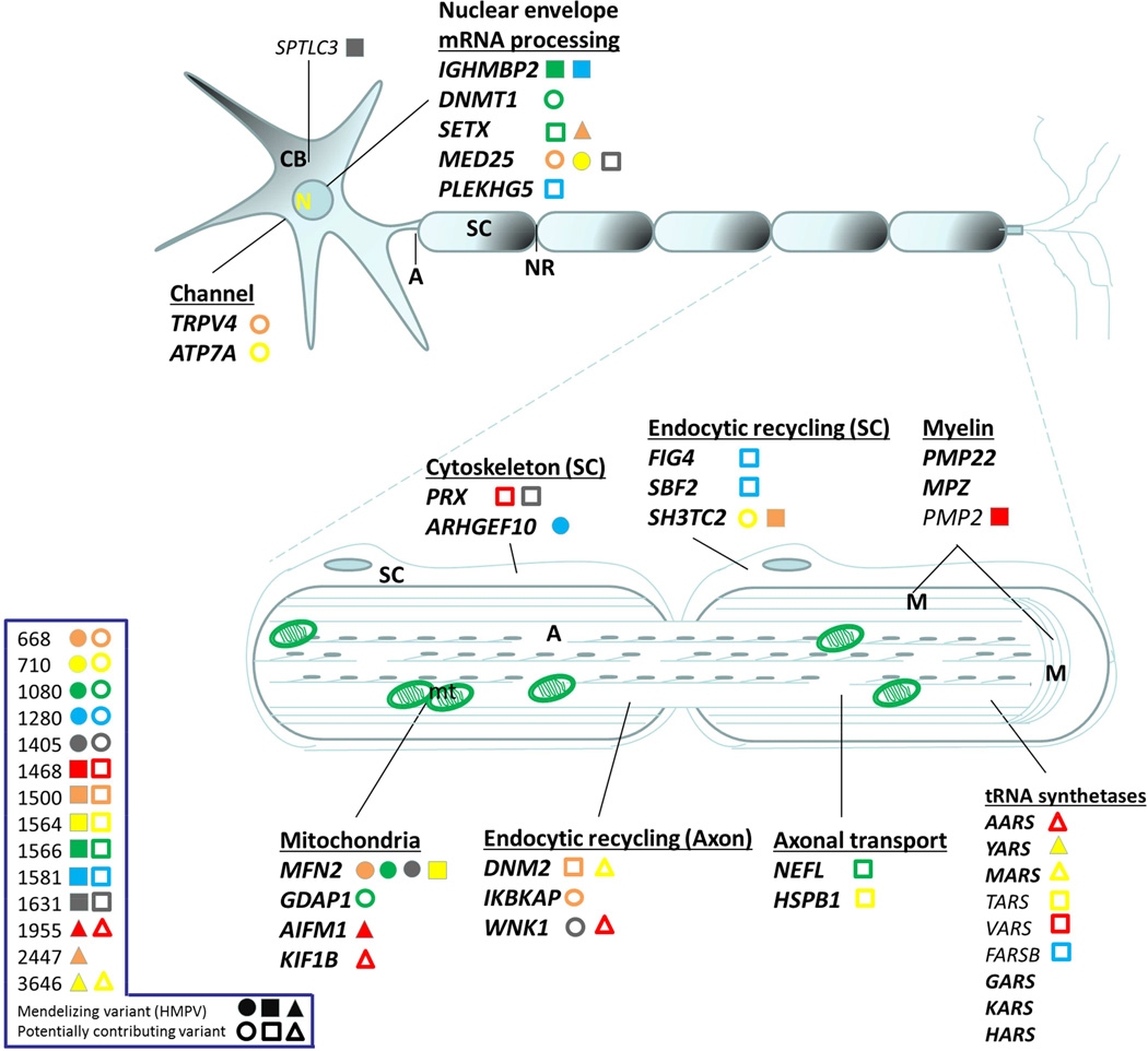
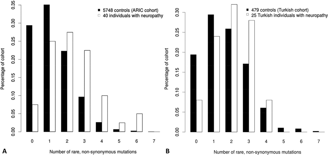
Charcot-Marie-Tooth (CMT) disease is a clinically and genetically heterogeneous distal symmetric polyneuropathy. Whole-exome sequencing (WES) of 40 individuals from 37 unrelated families with CMT-like peripheral neuropathy refractory to molecular diagnosis identified apparent causal mutations in ∼ 45% (17/37) of families. Three candidate disease genes are proposed, supported by a combination of genetic and in vivo studies. Aggregate analysis of mutation data revealed a significantly increased number of rare variants across 58 neuropathy-associated genes in subjects versus controls, confirmed in a second ethnically discrete neuropathy cohort, suggesting that mutation burden potentially contributes to phenotypic variability. Neuropathy genes shown to have highly penetrant Mendelizing variants (HPMVs) and implicated by burden in families were shown to interact genetically in a zebrafish assay exacerbating the phenotype established by the suppression of single genes. Our findings suggest that the combinatorial effect of rare variants contributes to disease burden and variable expressivity.


Figures

Figure 1
Figure 1
Pedigrees of CMT/neuropathy patients and segregation of causative mutations. A. Pedigree showing de novo occurrence of the known p.V244M MFN2 mutation in proband. B. Dominant pedigree of a dominant intermediate form of CMT and segregation of the identified novel variant p.E196Q in YARS. Mutation was inherited to the affected proband and affected sister from the affected mother. C. Pedigree of a dominant form of CMT and segregation of the mutation in candidate gene PMP2 (p.I43N). The affected proband inherited the mutation from his affected father, while both unaffected mother and sister do not carry the mutation. D. Pedigree of a dominantly inherited myopathy-neuropathy phenotype in a family with multiple affected individuals where a novel variant in DNAJB5 (p.P15S) was identified.
Figure 2
Figure 2
Suppression of pmp2 and sptlc3 in zebrafish causes defects in motor axon pathfinding and outgrowth. A–F. Lateral views of a control embryo, an embryo injected with pmp2 morpholino (MO) and embryos injected with pmp2 MO+PMP2_WT and pmp2 MO+PMP2_I43N, PMP2_WT and PMP2_I43N cocktails, respectively, at 2dpf (days post fertilization). Controls showed even spacing and normal branching of the motor neuron axons (A). In the pmp2 MO injected embryos the spacing of neuronal axons is perturbed by exiting the periphery but failing to extend (asterisks) or presenting pathfinding errors (arrows; B). Co-injection of pmp2 MO with human PMP2_WT resulted in restoration of the normal neuronal phenotype (C), but PMP2_I43N did not (D). Overexpression of human PMP2_WT causes mild pathfinding errors (E), suggesting dose sensitivity for PMP2. However, the human PMP2 mutant p.I43N, was significantly more severe than PMP2_WT when overexpressed (F) and had similar effects to suppression of pmp2 by MO knockdown. G. Percentage of normal versus abnormal embryos under the conditions being evaluated above. H–K. Wild type embryos (H) and sptlc3 morphants (I) in which secondary axons fail to migrate appropriately (white arrows). The phenotype induced by suppression of sptlc3 could be rescued by co-injection with SPTLC3_WT (J) but not SPTLC3_R150W (K). L. Quantification of normal embryos vs. embryos with motor neuron axon defects. For statistical analyses χ2 -tests were performed.
Figure 3
Figure 3
Neuron schematic of the localization or site of action of the main CMT/ neuropathy gene products. Legend on left shows patient identifier numbers and causative and possibly contributing mutations identified by WES. Full shapes correspond to rare presumed causative mutations deemed Highly Penetrant Mendelizing Variants (HMPVs); while empty shapes correspond to rare variants that may be contributing to the mutation burden in neuropathy patients. Each personal genome is distinguished by a unique color/shape. In bold are some of the canonical CMT genes.
Figure 4
Figure 4
Rare variant distribution in studied individuals suggests high carrier frequency for rare alleles in neuropathy genes in exome sequenced neuropathy cohort. A different extended cohort of 5748 Europeans from the ARIC-EA study was observed to have a tendency towards zero or one rare variants in recessive neuropathy genes.
Figure 5
Figure 5
Functional assessment of mutation burden hypothesis in a zebrafish model. First and second column panels show representative images of acetylated-tubulin (α-AcTub) staining of peripheral neurons in 2-day MO knockdown, single or in pair-wise combinations, zebrafish larvae. Third column panel shows qualitative assessment of morphant fish evaluated as defects in peripheral neuron axon extension, branching or pathfinding according to the scoring system developed. For pair-wise combinations, sub-effective concentrations of each of the gene-specific MOs were injected as shown in the graphs by the number of abnormal larvae in each category. However, when injected together increased severity in the phenotype was observed for all the pair-wise combinations, suggesting in vivo epistatic effects between these pairs of genes as observed in the α-AcTub fluorescence images and quantified in the graphs. Asterisks highlight some evidently affected axons. The scoring system used for assessing PNS defects in zebrafish was developed ad hoc and implemented here in order to best reflect the observations resulting from our experiments. Class I category refers to single axon defects; Class II category refers to two or more axons exhibiting defects with the presence of some normal axons; Class III category refers to generalized axonal defects; Class IV category refers to complete absence of axonal extension.
Figure 5
Figure 5
Functional assessment of mutation burden hypothesis in a zebrafish model. First and second column panels show representative images of acetylated-tubulin (α-AcTub) staining of peripheral neurons in 2-day MO knockdown, single or in pair-wise combinations, zebrafish larvae. Third column panel shows qualitative assessment of morphant fish evaluated as defects in peripheral neuron axon extension, branching or pathfinding according to the scoring system developed. For pair-wise combinations, sub-effective concentrations of each of the gene-specific MOs were injected as shown in the graphs by the number of abnormal larvae in each category. However, when injected together increased severity in the phenotype was observed for all the pair-wise combinations, suggesting in vivo epistatic effects between these pairs of genes as observed in the α-AcTub fluorescence images and quantified in the graphs. Asterisks highlight some evidently affected axons. The scoring system used for assessing PNS defects in zebrafish was developed ad hoc and implemented here in order to best reflect the observations resulting from our experiments. Class I category refers to single axon defects; Class II category refers to two or more axons exhibiting defects with the presence of some normal axons; Class III category refers to generalized axonal defects; Class IV category refers to complete absence of axonal extension.

References

    1. Allan W. Relation of hereditary pattern to clinical severity as illustrated by peroneal atrophy. Arch. Intern. Med. 1939;63:1123–1131.
    1. Anheim M, Tranchant C, Koenig M. The autosomal recessive cerebellar ataxias. N. Engl. J. Med. 2012;366(7):636–646. - PubMed
    1. Armstrong L, Biancheri R, Shyr C, Rossi A, Sinclair G, Ross CJ, Tarailo-Graovac M, Wasserman WW, van Karnebeek CD. AIMP1 deficiency presents as a cortical neurodegenerative disease with infantile onset. Neurogenetics. 2014;15(3):157–159. - PubMed
    1. Azzedine H, Senderek J, Rivolta C, Chrast R. Molecular genetics of charcot-marie-tooth disease: from genes to genomes. Mol Syndromol. 2012;3(5):204–214. - PMC - PubMed
    1. Ben Othmane K, Middleton LT, Loprest LJ, Wilkinson KM, Lennon F, Rozear MP, Stajich JM, Gaskell PC, Roses AD, Pericak-Vance MA, et al. Localization of a gene (CMT2A) for autosomal dominant Charcot-Marie-Tooth disease type 2 to chromosome 1p and evidence of genetic heterogeneity. Genomics. 1993;17(2):370–375. - PubMed

Publication types