. 2015 Jul 20:6:730.
doi: 10.3389/fmicb.2015.00730. eCollection 2015.

Reconstruction of the temporal signaling network in Salmonella-infected human cells

Affiliations

Reconstruction of the temporal signaling network in Salmonella-infected human cells

Gungor Budak et al. Front Microbiol. .

Abstract

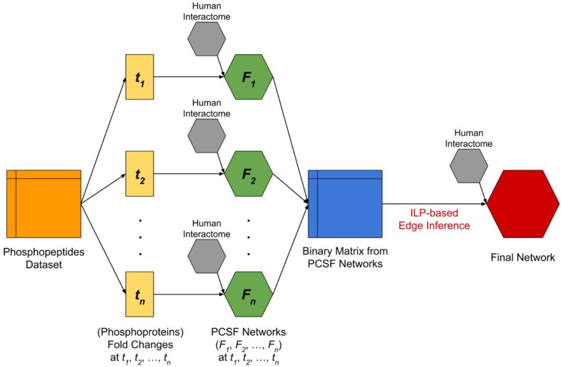
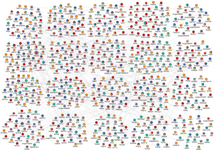
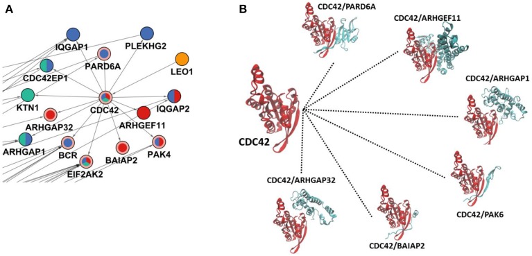
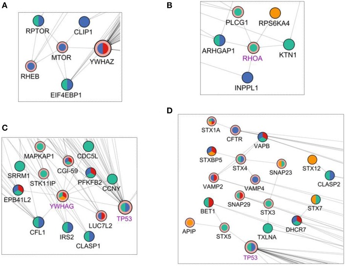
Salmonella enterica is a bacterial pathogen that usually infects its host through food sources. Translocation of the pathogen proteins into the host cells leads to changes in the signaling mechanism either by activating or inhibiting the host proteins. Given that the bacterial infection modifies the response network of the host, a more coherent view of the underlying biological processes and the signaling networks can be obtained by using a network modeling approach based on the reverse engineering principles. In this work, we have used a published temporal phosphoproteomic dataset of Salmonella-infected human cells and reconstructed the temporal signaling network of the human host by integrating the interactome and the phosphoproteomic dataset. We have combined two well-established network modeling frameworks, the Prize-collecting Steiner Forest (PCSF) approach and the Integer Linear Programming (ILP) based edge inference approach. The resulting network conserves the information on temporality, direction of interactions, while revealing hidden entities in the signaling, such as the SNARE binding, mTOR signaling, immune response, cytoskeleton organization, and apoptosis pathways. Targets of the Salmonella effectors in the host cells such as CDC42, RHOA, 14-3-3δ, Syntaxin family, Oxysterol-binding proteins were included in the reconstructed signaling network although they were not present in the initial phosphoproteomic data. We believe that integrated approaches, such as the one presented here, have a high potential for the identification of clinical targets in infectious diseases, especially in the Salmonella infections.

Keywords: Salmonella infection; network reconstruction; pathway analysis; phosphoproteomic; temporal data integration.


Figures

Figure 1
Figure 1
The flowchart of complete analysis. The dataset which includes temporal fold changes of phosphopeptides at four different time points (t1 = 2 min, t2 = 5 min, t3 = 10 min, t4 = 20 min) and at three different locations (nucleus, cytoplasm, and membrane) was split and converted into temporal fold changes datasets of the corresponding phosphoproteins by taking the maximum fold change among phosphopeptides that were observed at different locations and mapped to the same phosphoprotein. Next, we applied PCSF approach for each fold changes dataset by integrating human interactome in order to discover hidden intermediate proteins. The resulting networks (F1, F2, F3, F4) are then used to form a binary matrix where the rows are time points and columns are phosphoproteins. Each corresponding cell of the binary matrix represents a significant change (p < 0.05 and variance <15%) in the phosphoprotein at the time point. Finally, we applied an ILP-based edge inference approach by integrating human interactome in order to validate and determine edges and edge directions.
Figure 2
Figure 2
Visual representation of the reconstructed signaling network of Salmonella-infected host cell. The reconstructed network is clustered (Cluster #1 is at the left-top and Cluster #20 is at the right-bottom) and the layout of the network has been arranged accordingly for visualization. Time points when any node is found to be critically changed are shown with different colors on the nodes; yellow indicates a change at 2 min in the node, green indicates change at 5 min, blue indicates change at 10 min, and red indicates change at 20 min. If a node is present at more than one time points, then its coloring is a combination of colors of the corresponding time points. Steiner nodes differ from terminal nodes by a pink border and Salmonella—host interactome nodes differ from host nodes with a purple node label. Node sizes linearly proportional to the sum of in degree and out degree values of the nodes.
Figure 3
Figure 3
CDC42 and its interactions in the reconstructed network. (A) The region where CDC42 and its first neighbors are located in the reconstructed network. The coloring scheme is the same as in Figure 2 where CDC42, EIF2AK2, BCR, BAIAP2, ARHGAP32, PARD6A, and PAK4 are Steiner nodes and others are phosphoproteomic hits. (B) Structural details of CDC42 interactions where CDC42 uses the same binding site completely or partially to interact with its partners. Here, PAK6 is a structural homolog of PAK4; therefore to show similar binding of PAK4, it is represented with PAK6.
Figure 4
Figure 4
Visualization of the first degree neighbors of (A) mTOR, (B) RHOA, (C) YWHAG, and (D) Syntaxins in the reconstructed network in Figure 2. The coloring scheme is the same as in Figure 2.

References

    1. Aijo T., Granberg K., Lahdesmaki H. (2013). Sorad: a systems biology approach to predict and modulate dynamic signaling pathway response from phosphoproteome time-course measurements. Bioinformatics 29, 1283–1291. 10.1093/bioinformatics/btt130 - DOI - PubMed
    1. Auweter S. D., Yu H. B., Arena E. T., Guttman J. A., Finlay B. B. (2012). Oxysterol-binding protein (OSBP) enhances replication of intracellular Salmonella and binds the Salmonella SPI-2 effector SseL via its N-terminus. Microbes Infect. 14, 148–154. 10.1016/j.micinf.2011.09.003 - DOI - PubMed
    1. Bailly-Bechet M., Borgs C., Braunstein A., Chayes J., Dagkessamanskaia A., Francois J. M., et al. . (2010). Finding undetected protein associations in cell signaling by belief propagation. Proc. Natl. Acad. Sci. U.S.A. 108, 882–887. 10.1073/pnas.1004751108 - DOI - PMC - PubMed
    1. Bender C., Henjes F., Frohlich H., Wiemann S., Korf U., Beissbarth T. (2010). Dynamic deterministic effects propagation networks: learning signalling pathways from longitudinal protein array data. Bioinformatics 26, i596–i602. 10.1093/bioinformatics/btq385 - DOI - PMC - PubMed
    1. Berman H. M., Westbrook J., Feng Z., Gilliland G., Bhat T. N., Weissig H., et al. . (2000). The Protein Data Bank. Nucleic Acids Res. 28, 235–242. 10.1093/nar/28.1.235 - DOI - PMC - PubMed

LinkOut - more resources