. 2015 Aug 7:5:25.
doi: 10.1186/s13395-015-0051-9. eCollection 2015.

Involvement of adiponectin in the pathogenesis of dystrophinopathy

Affiliations

Involvement of adiponectin in the pathogenesis of dystrophinopathy

Michel Abou-Samra et al. Skelet Muscle. .

Abstract

Background: The hormone adiponectin (ApN) is decreased in the metabolic syndrome, where it plays a key pathogenic role. ApN also exerts some anti-inflammatory effects on skeletal muscles in mice exposed to acute or chronic inflammation. Here, we investigate whether ApN could be sufficiently potent to counteract a severe degenerative muscle disease, with an inflammatory component such as Duchenne muscular dystrophy (DMD).

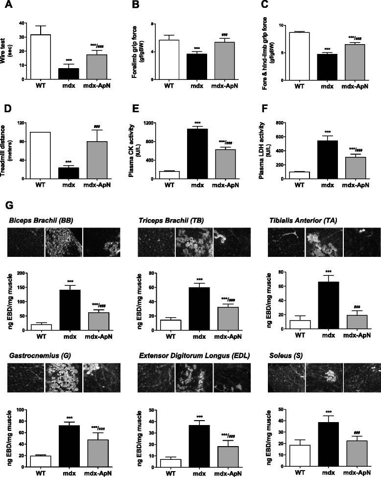
Methods: Mdx mice (a DMD model caused by dystrophin mutation) were crossed with mice overexpressing ApN in order to generate mdx-ApN mice; only littermates were used. Different markers of inflammation/oxidative stress and components of signaling pathways were studied. Global force was assessed by in vivo functional tests, and muscle injury with Evans Blue Dye (EBD). Eventually, primary cultures of human myotubes were used.

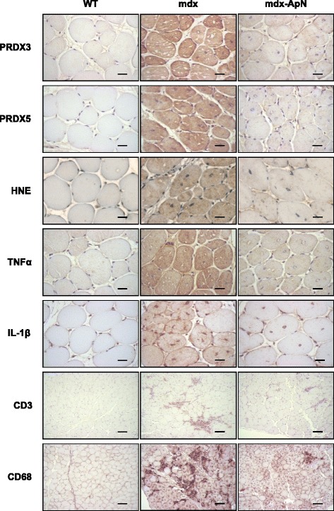
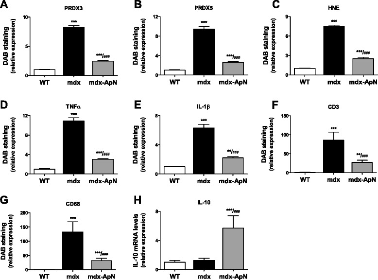
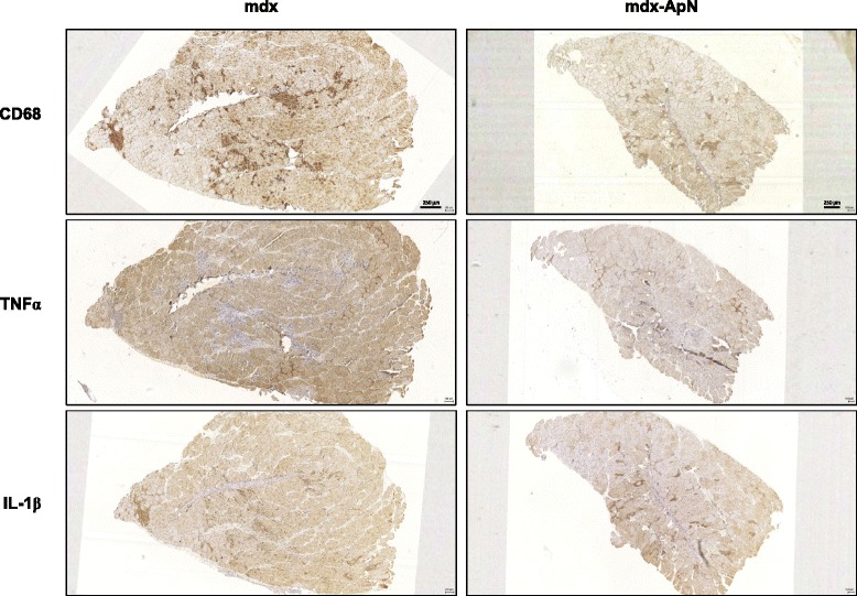
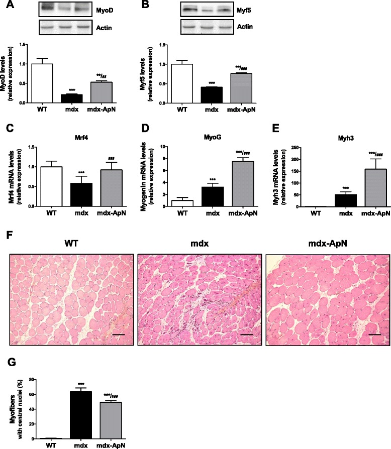
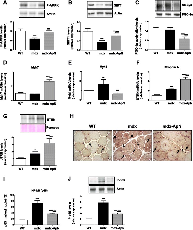
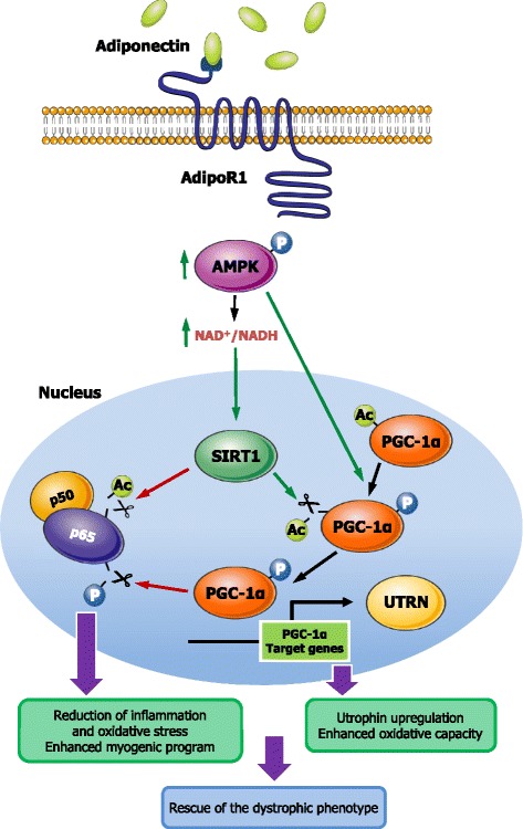
Results: Circulating ApN was markedly diminished in mdx mice. Replenishment of ApN strikingly reduced muscle inflammation, oxidative stress, and enhanced the expression of myogenic differentiation markers along with that of utrophin A (a dystrophin analog) in mdx-ApN mice. Accordingly, mdx-ApN mice exhibited higher global force and endurance as well as decreased muscle damage as quantified by curtailed extravasation of EBD in myofibers. These beneficial effects of ApN were recapitulated in human myotubes. ApN mediates its protection via the adiponectin receptor 1 (AdipoR1, the main ApN receptor in muscle) and the AMPK-SIRT1-PGC-1α signaling pathway, leading to downregulation of the nuclear factor kappa B (NF-κB) and inflammatory genes, together with upregulation of utrophin.

Conclusions: Adiponectin proves to be an extremely powerful hormone capable of protecting the skeletal muscle against inflammation and injury, thereby offering novel therapeutic perspectives for dystrophinopathies.

Keywords: AMPK signaling; Adiponectin; Inflammation; Mdx; NF-κB; Skeletal muscle; Utrophin.


Figures

Fig. 1
Fig. 1
Effects of adiponectin on different markers of oxidative stress and inflammation in muscles of mdx mice. Tibialis anterior muscles were sampled from 8-week-old WT mice, mdx mice, or mdx mice overexpressing adiponectin (mdx-ApN). Immunodetection was performed with specific antibodies directed against three oxidative stress markers (PRDX3/5, HNE), two pro-inflammatory cytokines (TNFα, IL-1β), one marker of T lymphocytes (CD3), and one macrophage marker (CD68). Representative sections for six mice per group are shown. Scale bar = 25 μm (for PRDX3/5, HNE, TNFα, and IL-1β) and 100 μm (for CD3 and CD68)
Fig. 2
Fig. 2
Quantification of markers of oxidative stress and inflammation in 8-week-old mdx mice. For each immunolabeling of Fig. 1 (a–g), the percentage of DAB deposit areas was calculated within muscle. h mRNA levels of IL-10, an anti-inflammatory cytokine. mRNA levels were normalized to cyclophilin. Data are means ± SD for six mice per group and the subsequent ratios were presented as relative expression compared to WT values. **p < 0.01; ***p < 0.001 vs. WT; ###p < 0.001 vs. mdx
Fig. 3
Fig. 3
Inflammation in the skeletal muscle of mdx mice. Successive sections of Tibialis anterior muscles from 8-week-old mdx and mdx-ApN mice were stained for CD68, TNFα, and IL-1β. Representative sections for six mice per group are shown. Scale bar = 250 μm
Fig. 4
Fig. 4
Effects of adiponectin on markers of the myogenic program in mdx mice. The expression of MyoD (a) and Myf5 (b), two myogenic regulatory factors, was analyzed by Western blotting in tibialis anterior muscles from the three groups of mice. Levels of each marker were normalized to actin levels. mRNA levels of Mrf4 (c) and myogenin (d), two markers of muscle differentiation. e mRNA levels of Myh3 (eMyHC), a marker of skeletal muscle regeneration. mRNA levels were normalized to cyclophilin and the subsequent ratios were presented as relative expression compared with WT values. f Tibialis anterior sections stained with hematoxylin-erythrosin-safran. Scale bar = 100 μm. g The percentage of muscle fibers with central nuclei was counted. Results are means ± SD; n = 6 mice per group. **p < 0.01; ***p < 0.001 vs. WT; ##p < 0.01; ###p < 0.001 vs. mdx mice
Fig. 5
Fig. 5
Effects of adiponectin on global force, resistance and muscle injury in mdx mice. Functional in vivo studies were carried out in mice from the three groups. a The animals were subjected to a wire test where they were suspended by their forelimbs and the time until they completely released the wire and fell down was recorded (seconds). b, c The mice were also lowered on a grid connected to a sensor to measure the muscle force of their forelimbs (b) or of both fore- and hindlimbs (c); data were then expressed in gram-force relative to body weight (gf/g BW). d Mice were submitted to a downhill treadmill exercise for 10 min during three consecutive days. On the 3rd session, the covered distance (meters) were measured for each mouse, 100 m being the maximal distance. Muscle injury was assessed by plasma activity of CK and LDH expressed as IU/L (basal state) (e, f) and by using EBD (g). Quantification of EBD extravasation was measured in different muscles after exercise. Six different muscles were sampled: biceps brachii, triceps brachii, gastrocnemius, tibialis anterior, extensor digitorium longus, and soleus. Qualitative detection of EBD was evaluated by fluorescence microscopy on frozen cryostat sections (see insets above histograms). Extravasated EBD concentrations were also quantified spectrophotometrically after extraction of the dye. Data were expressed as ng of EBD/mg muscle weight. The results presented herein are the means ± SD; n = 9 (a–c) and six (d–g) mice per group. ***p < 0.001 vs. WT ; ###p < 0.001 vs. mdx mice
Fig. 6
Fig. 6
Effects of adiponectin on AMPK signaling pathway and NF-κB activity in tibialis anterior muscles of mdx mice. The expression of P-AMPK (phosphorylated form) (a) and SIRT1 (b) was analyzed by Western blotting in muscles from the three groups of mice. c Densitometry of immunoprecipitation experiments performed on skeletal muscle lysates, using anti-PGC-1α antibody for precipitation and anti-acetyl-lysine antibody for immunoblotting. d mRNA levels of Myh7, a marker of slow twitch, oxidative myofibers. e mRNA levels of Myh1, a marker of fast twitch, glycolytic myofibers. f mRNA and g protein levels of utrophin A (UTRN) with a representative Western blot and Ponceau S stain. h Immunodetection of NF-κB (p65) in tibialis anterior sections; some positive marked nuclei (brown color) are indicated by arrows. Scale bar = 25 μm. i Quantification of p65 immunolabeling in myofiber nuclei (expressed as percent of total nuclei) in sections (as those shown in g). j Immunoblotting of phosphorylated NF-κB p65 in the same muscles. Levels of P-AMPK, SIRT1, and Acetyl-Lys were normalized to AMPK, actin, and PGC-1α levels, respectively. mRNA levels were normalized to cyclophilin, utrophin A protein levels to Ponceau, and P-p65 to Actin. The subsequent ratios were presented as relative expression compared to WT values. Results are means ± SD; n = 6 mice per group. *p < 0.05; **p < 0.01; ***p < 0.001 vs. WT; ###p < 0.001 vs. mdx mice
Fig. 7
Fig. 7
Effects of adiponectin on inflammatory markers in human skeletal muscle. TNFα (a) and IL-6 (b) mRNAs in primary culture of human myotubes: cells were treated with or without ApN, while being or not challenged with TNFα and IFNγ. TNFα (c) and IκB (d) mRNAs in human myotubes, which were transfected with siRNAs against AdipoR1, SIRT1, PGC-1α, or a negative (non-targeting, NT) control. After transfection, cells were treated with or without ApN, while being challenged with TNFα/IFNγ. mRNA levels were normalized to TATA box-binding protein. The subsequent ratios are presented as relative expression compared to control conditions [i.e., no TNFα/IFNγ and no ApN (a, b); NT siRNA without ApN (c, d)]. e Utrophin A mRNAs in human myotubes, which were treated with or without ApN, while being challenged with TNFα and IFNγ. mRNA levels were normalized as above. In this last graph, each patient is represented as being its own control (statistical analysis was performed on raw paired data). Data are means ± SD; n = 5–6 different subjects. **p < 0.01; ***p < 0.001 vs. no TNFα/IFNγ; ###p < 0.001 vs. TNFα/IFNγ without ApN; §§§p < 0.001 vs. all other conditions
Fig. 8
Fig. 8
Proposed model for the protective effects of adiponectin on dystrophic muscle. Signal transduction mediating ApN protection on dystrophic muscle: binding of ApN to AdipoR1 activates the AMPK/SIRT1/PGC-1α pathway. Briefly, ApN leads to AMPK phosphorylation/activation. P-AMPK in turn phosphorylates PGC-1α and indirectly increases the expression of SIRT1 (through rising NAD+/NADH ratio). SIRT1 in turn deacetylates and fully activates PGC-1α. Next, PGC-1α represses NF-κB activity by dephosphorylation of the p65 subunit [38], while SIRT1 represses it by deacetylation [39]. This results in decreased muscle inflammation/oxidative stress and improved myogenic program as well as enhanced utrophin expression and oxidative capacity, both processes helping rescue the dystrophic phenotype. Green arrow, stimulation; red arrow, inhibition

References

    1. Ouchi N, Walsh K. Adiponectin as an anti-inflammatory factor. Clin Chim Acta. 2007;380:24–30. doi: 10.1016/j.cca.2007.01.026. - DOI - PMC - PubMed
    1. Maury E, Brichard SM. Adipokine dysregulation, adipose tissue inflammation and metabolic syndrome. Mol Cell Endocrinol. 2010;314:1–16. doi: 10.1016/j.mce.2009.07.031. - DOI - PubMed
    1. Yamauchi T, Nio Y, Maki T, Kobayashi M, Takazawa T, Iwabu M, et al. Targeted disruption of AdipoR1 and AdipoR2 causes abrogation of adiponectin binding and metabolic actions. Nat Med. 2007;13:332–339. doi: 10.1038/nm1557. - DOI - PubMed
    1. Kadowaki T, Yamauchi T, Kubota N, Hara K, Ueki K, Tobe K. Adiponectin and adiponectin receptors in insulin resistance, diabetes, and the metabolic syndrome. J Clin Invest. 2006;116:1784–1792. doi: 10.1172/JCI29126. - DOI - PMC - PubMed
    1. Liu Y, Palanivel R, Rai E, Park M, Gabor TV, Scheid MP, et al. Adiponectin stimulates autophagy and reduces oxidative stress to enhance insulin sensitivity during high-fat diet feeding in mice. Diabetes. 2015;64:36–48. doi: 10.2337/db14-0267. - DOI - PubMed