

doi: 10.1155/2015/425438.
Epub 2015 Jul 15.
Extraction of Iron from the Rabbit Anterior Chamber with Reverse Iontophoresis
Affiliations
- PMID: 26257921
- PMCID: PMC4518192
- DOI: 10.1155/2015/425438
Item in Clipboard
Extraction of Iron from the Rabbit Anterior Chamber with Reverse Iontophoresis
J Ophthalmol.
2015.
Abstract
Ocular siderosis is a common eye disease caused by retention of an iron-containing intraocular foreign body in the eye. Iron-containing intraocular foreign bodies may cause severe inflammatory reaction and affect visual function. Currently the optimal treatment method of ocular siderosis is a moot point. This study used the reverse iontophoresis technique to noninvasively extract iron from the rabbit anterior chamber. By slit lamp observation and histological examination, reverse iontophoresis treatment has a good effect on ocular siderosis. Reverse iontophoresis seems to be a noninvasive and promising approach to extract iron from the anterior chamber to treat ocular siderosis.
Figures

Anterior segment photographs. (a) RI experiment cathode electrode and the eyecup. The cup is placed around eye cornea, and Ag/AgCl electrode is inserted into the eyecup to provide the cathode current for RI experiments. (b) The normal rabbit eye with smooth and transparent cornea, and transparent anterior chamber in the control group. (c) Immediate image after anterior chamber iron foreign body model. The anterior chamber was brown, and oozing around the pupil was observed. (d) 24 h after anterior iron foreign body model, there were conjunctival hyperemia, corneal edema, large bullous keratopathy, and rust-colored pigmentation, and the anterior chamber was brown. (e) Six days after modeling with 0 mA current RI. (f) Six days after modeling with 0.4 mA current RI.
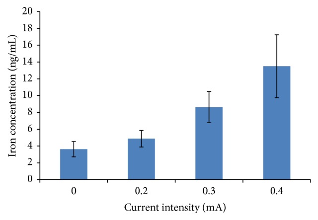
Iron concentrations of collected liquid following one-time RI with different current intensities.

Iron concentrations of collected liquid at different time points following RI.

Histological examination of cornea. Hematoxylin and eosin (H&E) staining of cornea from control group (a) and the iron-containing IOFB modeling group with 0 mA (b) and 0.4 mA RI treatment (c). Prussian blue iron staining of cornea from normal control group (d) and the iron-containing IOFB modeling group with 0 mA (e and f) and 0.4 mA RI treatment (g and h). (Original magnification, 20x. Scale bar, 50 μm.)
References
-
- Xie H., Chen S. Ocular siderosis. Eye science. 2013;28(2):108–112. - PubMed
-
- Duke-Elder S., MacFaul P. A. Injuries: I. Mechanical injuries. In: Duke-Elder S., editor. Systems of Ophthalmology. St. Louis, Mo, USA: Mosby; 1972. pp. 525–544.
LinkOut - more resources
Full Text Sources
Other Literature Sources
