Review
. 2015:2015:747543.
doi: 10.1155/2015/747543. Epub 2015 Jul 14.

Innate Immune Defenses in Human Tuberculosis: An Overview of the Interactions between Mycobacterium tuberculosis and Innate Immune Cells

Affiliations
Review

Innate Immune Defenses in Human Tuberculosis: An Overview of the Interactions between Mycobacterium tuberculosis and Innate Immune Cells

Jonathan Kevin Sia et al. J Immunol Res. 2015.

Abstract

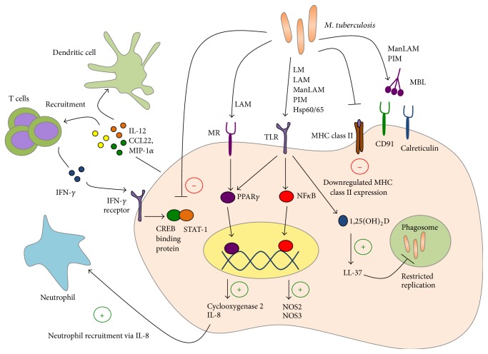
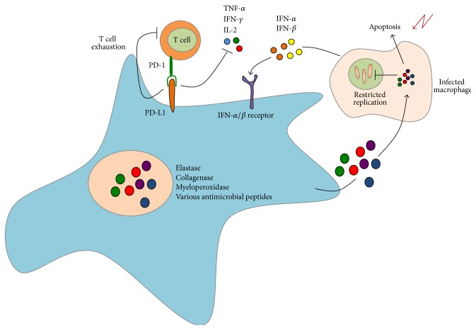
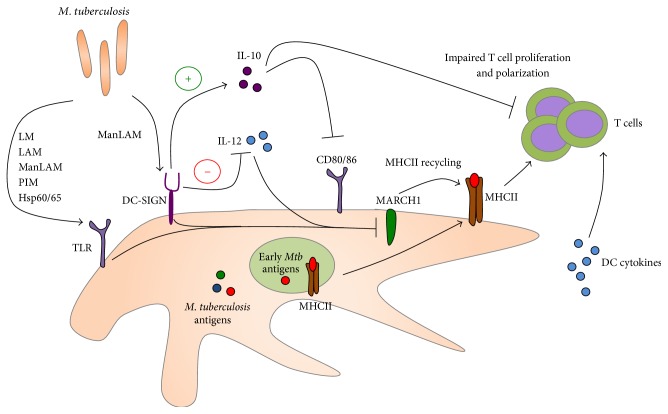
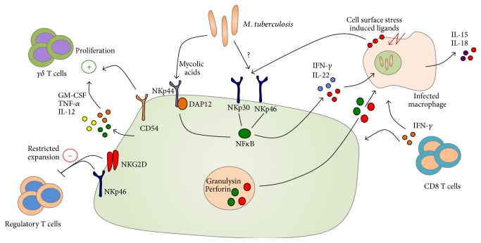
Tuberculosis (TB) remains a serious global public health problem that results in up to 2 million deaths each year. TB is caused by the human pathogen, Mycobacterium tuberculosis (Mtb), which infects primarily innate immune cells patrolling the lung. Innate immune cells serve as barometers of the immune response against Mtb infection by determining the inflammatory milieu in the lungs and promoting the generation of adaptive immune responses. However, innate immune cells are also potential niches for bacterial replication and are readily manipulated by Mtb. Our understanding of the early interactions between Mtb and innate immune cells is limited, especially in the context of human infection. This review will focus on Mtb interactions with human macrophages, dendritic cells, neutrophils, and NK cells and detail evidence that Mtb modulation of these cells negatively impacts Mtb-specific immune responses. Furthermore, this review will emphasize important innate immune pathways uncovered through human immunogenetic studies. Insights into the human innate immune response to Mtb infection are necessary for providing a rational basis for the augmentation of immune responses against Mtb infection, especially with respect to the generation of effective anti-TB immunotherapeutics and vaccines.


Figures

Figure 1
Figure 1
Human alveolar macrophages possess an array of receptors to recognize M. tuberculosis (Mtb). In response to Mtb infection, macrophages upregulate effector and signalling pathways to both prevent bacterial replication and recruit other immune cells into the site of infection. M. tuberculosis components, including lipoarabinomannans (LAMs), lipomannans (LMs), phosphatidylinositol mannosides (PIMs), and heat shock proteins (HSPs), are recognized by a variety of pattern recognition receptors. Following recognition of Mtb, host effectors, such as NF-κB and PPARγ, are activated to upregulate antimicrobial factors. These antimicrobial peptides (e.g., LL-37) possess both effector and signalling functions to actively interfere with bacterial replication as well as recruit and activate neutrophils, dendritic cells, and T cells. However, Mtb interferes with macrophage effector and signalling pathways. Most importantly, Mtb downregulates MHCII expression on macrophages to prevent optimal interaction with antigen specific T-cells. Furthermore, Mtb interferes with IFNγ signaling, a T cell cytokine mediator critical for upregulating the inherent antimicrobial capacity of macrophages during infection.
Figure 2
Figure 2
Neutrophils constitute a major subset of innate immune cells in the BAL and sputum of patients with active pulmonary TB. During infection with M. tuberculosis, neutrophils produce and secrete a variety of antimicrobial enzymes to restrict bacterial growth within infected macrophages. These neutrophil effectors promote apoptosis of infected macrophages, thereby limiting Mtb survival within infected host cells. However, these enzymes also mediate lung tissue damage and sustained, hyperactivated inflammatory response. Furthermore, transcriptional profiling studies have demonstrated the importance of PD-L1, a cell-surface associated molecule, in modulating T cell responses during infection with Mtb. Additional transcriptional studies have identified a blood based IFN-inducible gene signature in neutrophils that is unique to tuberculosis-specific immune responses.
Figure 3
Figure 3
DCs are the primary antigen presenting cells (APCs) in the immune system and play a central role in activation and differentiation of T cells by presenting antigenic peptides. DCs recognize a variety of M. tuberculosis components directly primarily through TLRs and DC-SIGN. Mtb impacts DC maturation and CD80/CD86 expression via induction of the immunosuppressive IL-10 mediator. Furthermore, engagement of TLRs and DC-SIGN during Mtb infection downregulates MARCH1, a ubiquitin ligase critical for recycling of MHCII on the cell surface. Downregulation of MARCH1 early during infection may play a role in limiting the repertoire of presented Mtb antigens and narrows the adaptive immune response in human TB.
Figure 4
Figure 4
Natural killer (NK) cells have the capacity to restrict M. tuberculosis replication through the production of soluble mediators such as GM-CSF, IL-12, TNF-α, IL-22, and IFN-γ. These upregulate the antimicrobial function of infected macrophages and activate antigen-specific T cell responses during M. tuberculosis infection. NK cell-derived antimicrobial factors such as granulysin and perforin indirectly restrict Mtb growth via the lysis of infected host cells. Several studies suggest that NK cells directly recognize Mtb-derived mycolic acids via NKp44. Aside from direct recognition of Mtb ligands, NKp30 and NKp46 recognize a variety of stress molecules upregulated on the surface of infected host cells.

References

    1. World Health Organization. Global Tuberculosis Report 2014. WHO; 2014.
    1. Dye C., Scheele S., Dolin P., Pathania V., Raviglione M. C. Global burden of tuberculosis: estimated incidence, prevalence, and mortality by country. WHO Global Surveillance and Monitoring Project. Journal of the American Medical Association. 1999;282(7):677–686. doi: 10.1001/jama.282.7.677. - DOI - PubMed
    1. Ronacher K., Joosten S. A., van Crevel R., Dockrell H. M., Walzl G., Ottenhoff T. H. M. Acquired immunodeficiencies and tuberculosis: focus on HIV/AIDS and diabetes mellitus. Immunological Reviews. 2015;264(1):121–137. doi: 10.1111/imr.12257. - DOI - PubMed
    1. Russell D. G. Who puts the tubercle in tuberculosis? Nature Reviews Microbiology. 2006;5(1):39–47. doi: 10.1038/nrmicro1538. - DOI - PubMed
    1. Saunders B. M., Cooper A. M. Restraining mycobacteria: role of granulomas in mycobacterial infections. Immunology and Cell Biology. 2000;78(4):334–341. doi: 10.1046/j.1440-1711.2000.00933.x. - DOI - PubMed

LinkOut - more resources