. 2015 Sep 3;525(7567):109-13.
doi: 10.1038/nature14670. Epub 2015 Aug 10.

Mutations in DCHS1 cause mitral valve prolapse

Affiliations

Mutations in DCHS1 cause mitral valve prolapse

Ronen Durst et al. Nature. .

Abstract

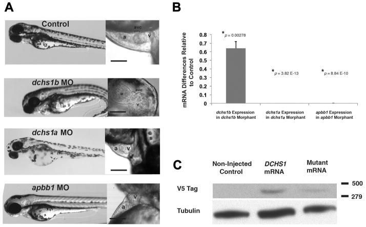
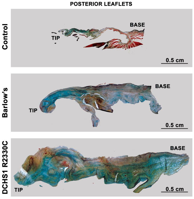
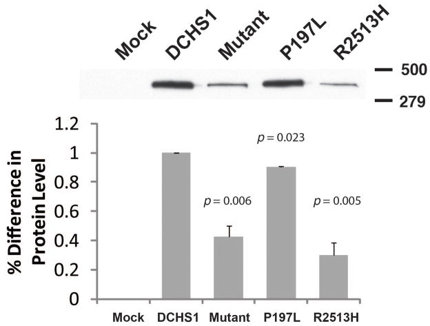
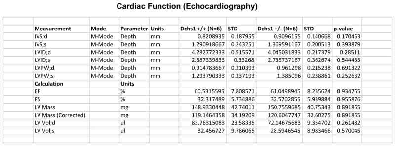
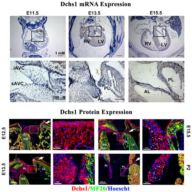
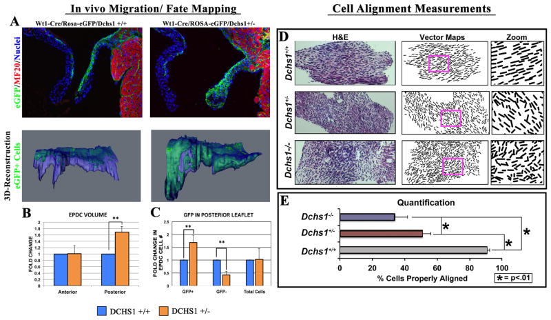
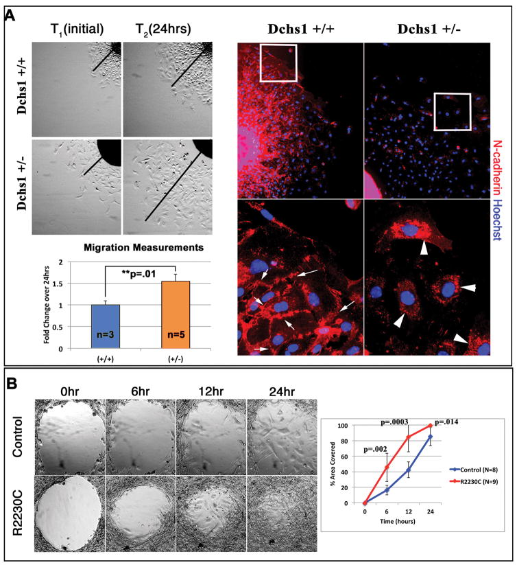
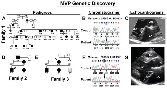
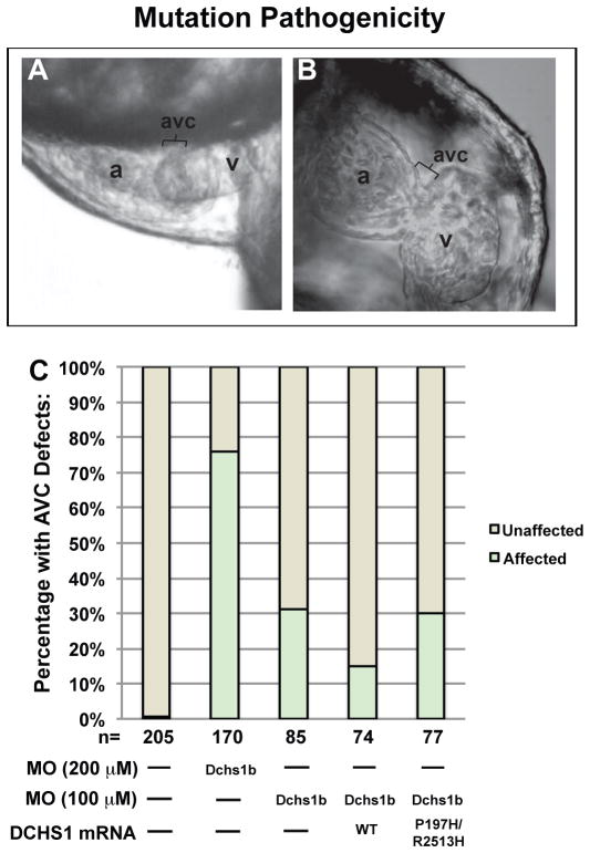
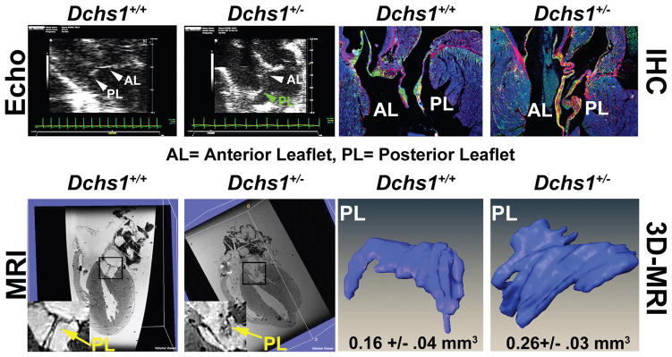
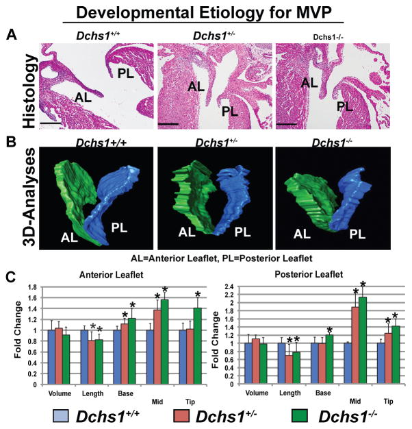
Mitral valve prolapse (MVP) is a common cardiac valve disease that affects nearly 1 in 40 individuals. It can manifest as mitral regurgitation and is the leading indication for mitral valve surgery. Despite a clear heritable component, the genetic aetiology leading to non-syndromic MVP has remained elusive. Four affected individuals from a large multigenerational family segregating non-syndromic MVP underwent capture sequencing of the linked interval on chromosome 11. We report a missense mutation in the DCHS1 gene, the human homologue of the Drosophila cell polarity gene dachsous (ds), that segregates with MVP in the family. Morpholino knockdown of the zebrafish homologue dachsous1b resulted in a cardiac atrioventricular canal defect that could be rescued by wild-type human DCHS1, but not by DCHS1 messenger RNA with the familial mutation. Further genetic studies identified two additional families in which a second deleterious DCHS1 mutation segregates with MVP. Both DCHS1 mutations reduce protein stability as demonstrated in zebrafish, cultured cells and, notably, in mitral valve interstitial cells (MVICs) obtained during mitral valve repair surgery of a proband. Dchs1(+/-) mice had prolapse of thickened mitral leaflets, which could be traced back to developmental errors in valve morphogenesis. DCHS1 deficiency in MVP patient MVICs, as well as in Dchs1(+/-) mouse MVICs, result in altered migration and cellular patterning, supporting these processes as aetiological underpinnings for the disease. Understanding the role of DCHS1 in mitral valve development and MVP pathogenesis holds potential for therapeutic insights for this very common disease.


Conflict of interest statement

Competing Interest Statement: The authors declare no competing financial interests

Figures

Extended Data Figure 1
Extended Data Figure 1. Measurement Of Endogenous And Exogenous Gene Expression In Danio Rerio
(A) Corresponding representative embryos of each morpholino knockdown on the left, with closeup of heart on the right. (B) To assess efficiency of morpholino knockdown, embryos were collected 72 hours after injection, mRNA was collected, and qPCR was performed. We demonstrate that morpholino (MO) knockdown of each indicated gene results in reduced mRNA expression, after normalization to beta-actin expression, compared to mock-injected controls (two-sided Student’s t-test). (C) Western blotting of embryos injected with DCHS1 mRNA demonstrates the production of protein. Mutant mRNA refers to the compound mutant P197L/R2513H. p-values are noted on graphs.
Extended Data Figure 2
Extended Data Figure 2. Apbb1 Is Not Expressed During Cardiac Morphogenesis
Apbb1 RNA expression was analyzed at E14.5 in sagittal sections using 2 separate antisense probes. Whereas strong cranial and neural expression is observed for Apbb1, no detectable cardiac expression or valve expression (arrow) is evident.
Extended Data Figure 3
Extended Data Figure 3. Dachsous1b Expression At The AV Junction
In situ hybridization reveals the presence of dchs1b in the AV canal (avc) at 54 hpf (A,B) and 72 hpf (C). The dchs1b expression is purple while a counterstain for cardiac tissue is brown (panel A). White arrows highlight the dchs1b signal in the AV canal.
Extended Data Figure 4
Extended Data Figure 4. Dachsous1b knockdown alters AV ring markers
In situ hybridization at 48hpf and 72hpf, as indicated, was performed for known AV ring markers. In contrast to wt (A) bmp4 expression is expanded into the ventricle at 48 hpf in dchs1 knockdown embryos at 48 hpf (B), spp1 and notch1b expression was largely unperturbed (C–F), and has2 expression was not detected at 48 hpf, and is faint at 72 hpf in dchs1 knockdown, compared to identically handled and stained controls (I–L).
Extended Data Figure 5
Extended Data Figure 5. Histopathology Mitral Valves
Human posterior leaflets of control, Barlow’s with MVP, and DCHS1 p.R2330C were isolated, fixed and stained with Movats Pentachrome. Leaflet thickening, elongation and myxomatous degeneration is observed in the Barlow’s and DCHS1 p.R2330C leaflets compared to controls. Expansion of the proteoglycan layer (blue) and disruption of the normal stratification of matrix boundaries is observed in the Barlow’s and DCHS1 p.R2330C leaflets. Blue=proteoglycan, Yellow=collagen, Black=elastin, Red=fibrin or cardiac muscle. Scale bars = 0.5cm
Extended Data Figure 6
Extended Data Figure 6. Protein Expression Of Uncoupled Mutations
In order to determine which Family 1 DCHS1 mutation is leading to the observed decrease in protein expression, constructs were generated that harbored only the p.P197L or the p.R2513H variant. Mutant refers to the double mutant P197L/R2513H construct. Western blot analyses from transfected HEK293 cells demonstrate that the p.R2513H mutation causes a significant decrease in DCHS1 protein expression, similar to that of the construct with both variants (mutant), suggesting pathogenicity. Percent difference in protein levels is depicted. Normalization of data was accomplished by qPCR specific to the transfected constructs. p-values are denoted in graphs.
Extended Data Figure 7
Extended Data Figure 7. Cardiac Function Is Not Altered In Dchs1+/− Mice
M-Mode analyses were performed to determine whether cardiac structure and/or function were perturbed in the Dchs1+/− mice. No statistically significant differences were observed in either cardiac structure or calculated cardiac function (N=6 for each genotype). Abbreviations: IVS-interventricular septum, d-diastole, s-systole, LVID-left ventricular internal dimension, LVPW-left ventricular posterior wall, EF-ejection fraction, FS-fractional shortening, LV-left ventricle
Extended Data Figure 8
Extended Data Figure 8. Dchs1 Expression During Cardiac Development
Upper Panel: RNA expression of Dchs1 was analyzed during embryonic gestation (E11.5, E13.5, and E15.5) by section in situ hybridization. At E11.5 Dchs1 RNA (blue staining) expression is observed in the endocardium and mesenchyme of the superior and inferior cushions (sAVC and iAVC, respectively). A gradient pattern of expression is observed at this time point with more intense expression near the endocardium. At E13.5 and E15.5, a similar pattern is observed in the forming anterior and posterior mitral leaflets (AL and PL, respectively). Lower Panel: Dchs1 protein expression (red) is observed throughout cardiac development in the endothelial cells and interstitial cells of the developing valves. Dchs1 shows asymmetric expression in the valvular interstitial cell bodies around E15.5 (arrowheads). Dchs1 protein is also observed in the epicardium and AV sulcus (arrows). (Red-Dchs1, Green-MF20, Blue-Hoescht).
Extended Data Figure 9
Extended Data Figure 9. Dchs1 Deficiency Causes Altered Valvular Interstitial Cell Patterning In Vivo.
(A) IHC for eGFP of postnatal day 0 (P0) lineage traced Wt1-Cre/Rosa-eGFP/Dchs1+/+ neonatal mice show epicardial-derived cells (EPDCs) migrating into the posterior leaflet as a sheet of cells directly under the endothelium of the atrialis. This normal patterning is perturbed in the Wt1-Cre/Rose-eGFP/Dchs1+/− mice. 3D reconstructions were used to examine all EPDCs in the posterior leaflet of both genotypes to obtain a complete fate map of these cells. (B, C) Total volume of the leaflet is unchanged at this time point. However, the total volume of EPDCs as well as total EPDC cell number is significantly increased. There is a significant decrease in the number of non-EPDCs in the posterior leaflet with no overall change in total cell number. These data demonstrate that a minimum threshold of Dchs1 expression is required for normal migration of EPDCs into the posterior leaflet, normal patterning of this cell population, and cross-talk between EPDC and non-EPDC cell types in the valve. (**p<.01) (D) Isolated anterior mitral leaflet from fetal (E17.5) Dchs1+/+, Dchs1+/−, and Dchs1−/− mice were used to quantify cellular alignment of valvular interstitial cells. Vector maps were generated from histological (H&E) stains to show orientation and alignment of cells in relationship to each other. Boxes in each vector map panel are represented as zoomed images of regions within each of the valves to show cell orientation. (E) Cell alignment and polarity were quantified as the number of cells that deviate >10 degrees from the proximal-distal (P–D) axis of the leaflet. 90% of the cells in Dchs1+/+ show proper alignment with each other and along this P–D axis. Haploinsufficiency (Dchs1+/−) results in a 50% reduction in cell alignment, which is further reduced in Dchs1−/− (*=p-values<.01).
Extended Data Figure 10
Extended Data Figure 10. Mice And MVP Patients With Dchs1 Deficiency Exhibit Migratory Defects In Vitro
(A) Posterior Leaflets of P0 neonatal Dchs1+/+ and Dchs1+/− mice were explanted and interstitial cells were allowed to migrate out for 24 hours. Dchs1+/− mice exhibit increased migration (black lines drawn from explants) coincident with loss of cell-cell contacts and N-Cadherin expression at focal adhesions. Whereas N-Cadherin expression (red) is found at the membrane at points of cell-cell contract in Dchs1+/+ valvular interstitial cells (arrows), this membrane expression is lost in the Dchs1+/− cells and is prominently expressed in the cytoplasm (arrows). (Nuclei-blue). (B) Migration assays using control and MVP patient (p.R2330C) valvular interstitial cells exhibit a similar affect as observed in the mouse cells whereby the p.R2330C cells exhibit an increase in migration. p-values are denoted in graphs.
Figure 1
Figure 1. Pedigrees, mutation, and phenotype
Black symbols = MVP affecteds, gray = unknown, Arrows = probands, If no genotype is shown, the individuals were unavailable for study. (A). Pedigree linked to chromosome 11. #= Individuals under 15 years of age, *- individuals sequenced. Genotypes c. 7538G>A (R2513H) of DCHS1 mutation are shown. (B) DNA sequence of c.7538G>A (p.R2513H). (C) Two-dimensional echocardiographic long-axis view of Family 1 proband. Dashed line marks mitral annulus. (D,E) Family 2 and 3 pedigrees. Genotype c.6988C>T (p.R2330C) shown. (F) DNA sequence c. 6988C>T (p.R2330C) (G) Two-dimensional echocardiographic long-axis view of Family 2 proband.
Figure 2
Figure 2. Zebrafish Dchs1b Is Required For AV Canal Development
(A) By 72 hpf, zebrafish hearts develop a constriction in the atrioventricular canal (AVC) that separates the atrium (a) from the ventricle (v). (B) Knockdown of dchs1b results in absence of the AV constriction (bracket). (C) ~75% of Dchs1b morphants exhibit AVC defects (*p= 1×10−62). DCHS1 human mRNA rescues the dchs1b morpholino AVC phenotype whereas human mutant DCHS1 mRNA (P197H/R2513H) fails to rescue (**p=.009). Total number of fish analyzed was 611 and statistical values were obtained using Fisher’s Exact test.
Figure 3
Figure 3. DCHS1 Mutations Result In Diminished Protein Levels
(A–C) Western blot: (p.P197L/p.R2513H) mutant DCHS1 results in a 60% decrease in protein with no change in RNA expression. (D) Left Panel: DCHS1 wild-type (WT) or mutant (p.P197L/p.R2513H) transfectants treated with cycloheximide for specified times followed by Western analyses. Right Panel: Cycloheximide on Control (DCHS1 WT) or MVP patient (p.R2330C) MVICs. Tubulin=loading control (E) Graphical depiction of calculated protein half-lives. WT and mutant transfectants half-life = 5.8 hours versus 1.6, respectively (left graph). Control and mutant DCHS1 half-life is 1.73 hours versus 0.46 hours, respectively (right graph). Analyses performed in triplicate and repeated four times. Error bars = standard deviations; p-values calculated using two-tailed Students t-test.
Figure 4
Figure 4. Dchs1 Deficiency Causes MVP and Myxomatous Degeneration in the Adult Mouse
Echocardiography (Echo), MRI, Histopathology, and 3D reconstructions performed on 9-month old male Dchs1+/+ and Dchs1+/− mouse hearts. Echo: posterior leaflet prolapse in Dchs1+/− (green arrow) (N=6/genotype). Immunohistochemistry (IHC): Dchs1+/− (N=5) anterior, posterior leaflets (AL, PL) exhibit myxomatous degeneration and expansion of proteoglycan expression compared to Dchs1+/+ (N=7), collagen (red), proteoglycans (green). MRI show posterior leaflet (PL) thickening in Dchs1+/− (arrow-inset) compared to control littermates. 3D reconstructions of MRI: Dchs1+/− mice exhibit thickened and elongated leaflets compared to Dchs1+/+. (Two-tailed Student t-test was used to calculate p-values; p=.01, N=4/genotype).
Figure 5
Figure 5. Developmental Etiology for MVP
(A,B) H&E and 3D reconstructions of E17.5 Dchs1+/+, Dchs1+/−, and Dchs1−/− mouse hearts showing thickening of anterior and posterior leaflets (AL, PL) in Dchs1−/− mice compared to Dchs1+/+. Dchs1+/− valves display an intermediate phenotype. (C) Quantification of valve dimensions showing Dchs1−/− (green bars) and Dchs1+/− (orange bars) anterior and posterior lengths were significantly reduced compared to Dchs1+/+ (blue bars) leaflets. Dchs1−/− and Dchs1+/− valves displayed increased thickness throughout the leaflets compared to Dchs1+/+. Scale Bars= 100μm. N=5/genotype and two-tailed Student’s t-test was used to calculate p-values; *p<.01

References

    1. Freed LA, et al. Mitral valve prolapse in the general population: the benign nature of echocardiographic features in the Framingham Heart Study. J Am Coll Cardiol. 2002;40:1298–1304. S0735109702021617 [pii] - PubMed
    1. Avierinos JF, et al. Natural history of asymptomatic mitral valve prolapse in the community. Circulation. 2002;106:1355–1361. - PubMed
    1. Freed LA, et al. Prevalence and clinical outcome of mitral-valve prolapse. N Engl J Med. 1999;341:1–7. doi: 10.1056/NEJM199907013410101. - DOI - PubMed
    1. Vaishnava P, Fuster V, Goldman M, Bonow RO. Surgery for asymptomatic degenerative aortic and mitral valve disease. Nature reviews. Cardiology. 2011;8:173–177. doi: 10.1038/nrcardio.2010.203. - DOI - PubMed
    1. Waller BF, Maron BJ, Del Negro AA, Gottdiener JS, Roberts WC. Frequency and significance of M-mode echocardiographic evidence of mitral valve prolapse in clinically isolated pure mitral regurgitation: analysis of 65 patients having mitral valve replacement. The American journal of cardiology. 1984;53:139–147. - PubMed

Publication types

MeSH terms