. 2015 Sep;11(9):697-704.
doi: 10.1038/nchembio.1866. Epub 2015 Aug 10.

An excess of catalytically required motions inhibits the scavenger decapping enzyme

Affiliations

An excess of catalytically required motions inhibits the scavenger decapping enzyme

Ancilla Neu et al. Nat Chem Biol. 2015 Sep.

Abstract

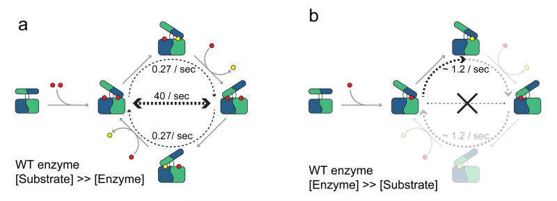
The scavenger decapping enzyme hydrolyzes the protective 5' cap structure on short mRNA fragments that are generated from the exosomal degradation of mRNAs. From static crystal structures and NMR data, it is apparent that the dimeric enzyme has to undergo large structural changes to bind its substrate in a catalytically competent conformation. Here we studied the yeast enzyme and showed that the associated opening and closing motions can be orders of magnitude faster than the catalytic turnover rate. This excess of motion is induced by the binding of a second ligand to the enzyme, which occurs at high substrate concentrations. We designed a mutant that disrupted the allosteric pathway that links the second binding event to the dynamics and showed that this mutant enzyme is hyperactive. Our data reveal a unique mechanism of substrate inhibition in which motions that are required for catalytic activity also inhibit efficient turnover when they are present in excess.


Figures

Figure 1
Figure 1. Structure of the yeast DcpS:substrate complex
(a) 2.3 Å resolution crystal structure of the Dcs1p enzyme from S. cerevisiae in complex with m7GDP. The dimeric enzyme adopts an asymmetric conformation with one closed and one open binding site. The protein chains are coloured blue with magenta side chains and green with yellow side chains respectively and the substrate is coloured red. The boxed region is shown in panel b in a similar orientation. A cartoon representing the conformation of the enzyme is indicated, where the red dot refers to the substrate. (b) The substrate is tightly bound in the closed binding site. Residues Y94 and W161 highlight the high asymmetry of the enzyme in the ligand bound form; these aromatic rings come within 5 Å of each other in the closed binding site, but are more than 22 Å apart in the open binding site. Without conformational changes, the substrate (product) is not able to dissociate from the enzyme.
Figure 2
Figure 2. The scavenger decapping enzyme binds two substrates in a sequential manner
(a) Methyl-TROSY NMR spectrum of the free, symmetric enzyme (0.25 mM dimer concentration). Assignments for a number of isoleucine and methionine residues that are close to the active site are indicated (see also Supplementary Fig. 2). Cartoons indicate the state of the complex. (b) Methyl-TROSY NMR spectrum of Dcs1p after addition of one molar equivalent of m7GpppG (0.25 mM) to the dimeric enzyme. A number of resonances that report on the symmetric to asymmetric conversion are indicated with arrows. (c) As in (b), but after addition of 20 molar equivalents of substrate (5 mM). Resonances that result from residues in the open binding site shift, indicating that a second substrate interacts in the open binding site of the enzyme. The insets on the right show a detailed view of the sequential binding process. (d) Plot of the changes in the peak intensities for residue I12 (open and closed resonances) and the changes in the peak position for residue M153 (CSP in carbon) for 15 titrations points (see a-c and boxed regions in the spectra of panels b and c). The errors (s.d.) in the extracted binding constants are based on Monte Carlo simulations for a simultaneous fit of multiple residues (Supplementary Fig. 3a). For clarity the scale of the x-axis is shown different below and above the 1:1 m7GpppG:Dcs1p ratio, as is indicated by the waveform.
Figure 3
Figure 3. Quantification of domain flipping motions in the scavenger decapping enzyme
(a) Longitudinal exchange experiment that directly reports on motions in the enzyme where the open binding site closes and where the closed binding site opens. The cyan and pink coloured resonances result from the closed and open sites, respectively. The arrows point to the resonances that appear due to the domain flipping motions. The spectrum shown here is recorded with a mixing time of 75 ms in the presence of a 20-fold excess of substrate over the dimeric enzyme. (b) Quantification of the exchange process in the presence of a 20 fold excess of ligand. The circles indicate resonance intensities of the cross and auto peaks. The error bars represent uncertainties in the resonance intensities. The drawn lines are a best global fit to the data and yield an exchange rate of 35.4 (± 4.8) s−1. The error (s.d.) in the extracted parameters are based on 100 Monte Carlo simulations. (c) The flipping motions (a, b) depend on the m7GpppG ligand excess. Higher excess of ligand results in a higher occupation of the open second binding site (Figure 2c, d) and in faster exchange rates. This correlation between exchange rates and binding site occupancy shows that the flipping motions are directly induced by the presence of the second ligand in the open binding site.
Figure 4
Figure 4. Turnover rates of the enzyme are much slower than the flipping rates
(a) 31P NMR spectra that report on the Dcs1p mediated degradation (50 nM enzyme) of the (0.5 mM) m7GpppG substrate (red, top) into m7GMP and GDP product (bottom, yellow). NMR spectra (18 minutes each) were recorded successively until completion of the reaction. (b) Progression curves of the reaction. Crosses correspond to the mean concentration of the substrate and product signals and are derived from the peak intensities of all 31P signals. The drawn line corresponds to the best fit of the data, the extracted turnover rates are indicated. (c) Longitudinal exchange experiment of Dcs1p K126A in the presence of a 20 fold excess of substrate and using a mixing time of 75 ms (where the exchange peaks have maximum intensity for the WT protein). Exchange peaks were not observed (arrows in the traces) demonstrating that the K126A enzyme does not undergo unproductive flipping motions.
Figure 5
Figure 5. Cartoon representation of the substrate inhibition mechanism of the scavenger decapping enzyme
(a) Under substrate excess, both binding sites in the enzyme are occupied. This results in fast non-productive flipping motions (horizontal dashed arrow) and low turnover rates (curved dashed arrows). Substrate and product is indicated with red and yellow circles respectively. Determined rates are indicated. (b) Under single turnover conditions, only one of the two binding sites in the enzyme is occupied. The unproductive motions (crossed out horizontal line) no longer take place and the catalytic turnover is increased (curved dashed arrow). The indicated turnover rate corresponds to the human enzyme and may differ for the yeast complex. Note that under these conditions part of the catalytic cycle is not sampled and indicated in a light colour for reference only.

References

    1. Fersht AR. Structure and mechanism in protein science: A guide to enzyme catalysis and protein folding. W. H. Freeman; 1999.
    1. Hammes-Schiffer S, Benkovic SJ. Relating protein motion to catalysis. Annu Rev Biochem. 2006;75:519–41. - PubMed
    1. Boehr DD, Dyson HJ, Wright PE. An NMR perspective on enzyme dynamics. Chem Rev. 2006;106:3055–79. - PubMed
    1. Berman HM, et al. The Protein Data Bank. Nucleic Acids Res. 2000;28:235–42. - PMC - PubMed
    1. Henzler-Wildman K, Kern D. Dynamic personalities of proteins. Nature. 2007;450:964–72. - PubMed

Publication types

MeSH terms

Associated data

LinkOut - more resources