Review
. 2015 Aug 6;7(8):4438-60.
doi: 10.3390/v7082829.

RNA-Dependent RNA Polymerases of Picornaviruses: From the Structure to Regulatory Mechanisms

Affiliations
Review

RNA-Dependent RNA Polymerases of Picornaviruses: From the Structure to Regulatory Mechanisms

Cristina Ferrer-Orta et al. Viruses. .

Abstract

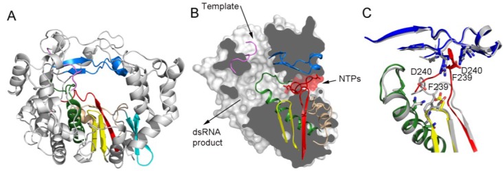
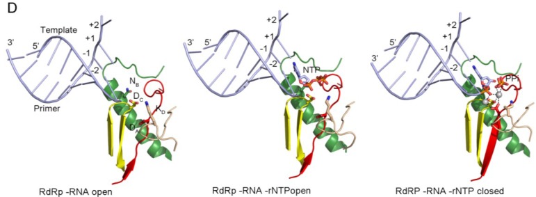
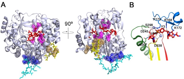
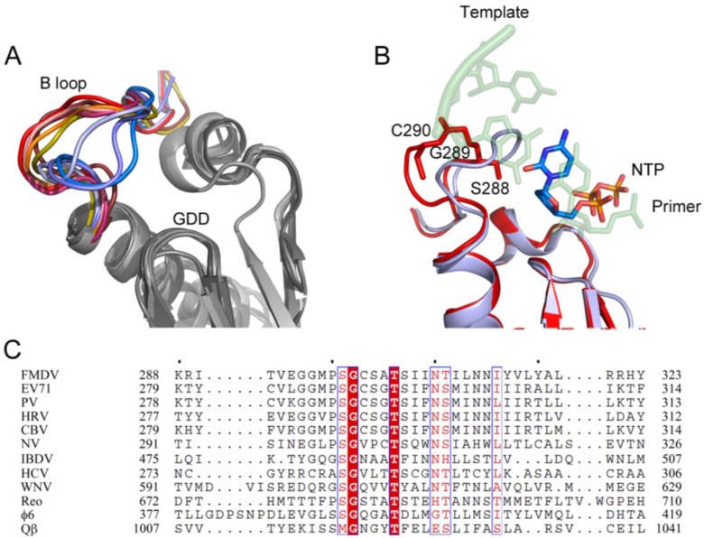
RNA viruses typically encode their own RNA-dependent RNA polymerase (RdRP) to ensure genome replication within the infected cells. RdRP function is critical not only for the virus life cycle but also for its adaptive potential. The combination of low fidelity of replication and the absence of proofreading and excision activities within the RdRPs result in high mutation frequencies that allow these viruses a rapid adaptation to changing environments. In this review, we summarize the current knowledge about structural and functional aspects on RdRP catalytic complexes, focused mainly in the Picornaviridae family. The structural data currently available from these viruses provided high-resolution snapshots for a range of conformational states associated to RNA template-primer binding, rNTP recognition, catalysis and chain translocation. As these enzymes are major targets for the development of antiviral compounds, such structural information is essential for the design of new therapies.

Keywords: RNA-dependent RNA polymerase; picornaviruses; positive-strand RNA viruses; replication fidelity; viral replication.


Figures

Figure 1
Figure 1
Overall structure of a viral RdRP. (A) Ribbon representation of a typical picornaviral RdRP (model from the cardiovirus EMCV 3Dpol, PDB id. 4NZ0). The seven conserved motifs are indicated in different colours: motif A, red; motif B, green; motif C, yellow; motif D, sand; motif E, cyan; motif F, blue; motif G, pink; (B) Lateral view of a surface representation of the enzyme (grey) that has been cut to expose the three channels that are the entry and exit sites of the different substrates and reaction products. The structural elements that support motifs A–G are also shown as ribbons. This panel also shows the organization of the palm sub-domain with motif A shown in two alternative conformations: the standard conformation (PDB id. 4NZ0) found in the apo-form of most crystallized 3Dpol proteins and the altered conformation found int the tetragonal crystal form of the EMCV enzyme (PDB id. 4NYZ). The alterations affect mainly Asp240, the amino acid in charge of incoming ribonucleotide triphosphate (rNTP) selection, and the neighboring Phe239 that move ~10 Å away from its position in the enzyme catalytic cavity directed towards the entrance of the nucleotide channel, approaching to motif F; (C) Close up of the structural superimposition of the two alternative conformations of the EMCV motif A; (D) The PV replication-elongation complexes. Sequential structures illustrating the movement of the different palm residues from a binary PV 3Dpol-RNA open complex (left) to an open 3Dpol-RNA-rNTP ternary complex (middle) where the incoming rNTP is positioned in the active site for catalysis and, a closed ternary complex (right) after nucleotide incorporation and pyrophosphate (PPi) release. The residues DA (involved in rNTP selection through an interaction with the 2′ hydroxyl group), DC (the catalytic aspartate of motif C), KD (the general acid residue of motif D that can coordinate the export of the PPi group) and NB (a conserved Asn of motif B, interacting with DA) have been highlighted as sticks. The different structures correspond to the 3Dpol-RNA (PDB id. 3OL6), 3Dpol-RNA-CTP open complex (PDB id. 3OLB) and 3Dpol-RNA-CTP closed complex (PDB id. 3OL7) structures of PV elongation complexes, respectively [7].
Figure 1
Figure 1
Overall structure of a viral RdRP. (A) Ribbon representation of a typical picornaviral RdRP (model from the cardiovirus EMCV 3Dpol, PDB id. 4NZ0). The seven conserved motifs are indicated in different colours: motif A, red; motif B, green; motif C, yellow; motif D, sand; motif E, cyan; motif F, blue; motif G, pink; (B) Lateral view of a surface representation of the enzyme (grey) that has been cut to expose the three channels that are the entry and exit sites of the different substrates and reaction products. The structural elements that support motifs A–G are also shown as ribbons. This panel also shows the organization of the palm sub-domain with motif A shown in two alternative conformations: the standard conformation (PDB id. 4NZ0) found in the apo-form of most crystallized 3Dpol proteins and the altered conformation found int the tetragonal crystal form of the EMCV enzyme (PDB id. 4NYZ). The alterations affect mainly Asp240, the amino acid in charge of incoming ribonucleotide triphosphate (rNTP) selection, and the neighboring Phe239 that move ~10 Å away from its position in the enzyme catalytic cavity directed towards the entrance of the nucleotide channel, approaching to motif F; (C) Close up of the structural superimposition of the two alternative conformations of the EMCV motif A; (D) The PV replication-elongation complexes. Sequential structures illustrating the movement of the different palm residues from a binary PV 3Dpol-RNA open complex (left) to an open 3Dpol-RNA-rNTP ternary complex (middle) where the incoming rNTP is positioned in the active site for catalysis and, a closed ternary complex (right) after nucleotide incorporation and pyrophosphate (PPi) release. The residues DA (involved in rNTP selection through an interaction with the 2′ hydroxyl group), DC (the catalytic aspartate of motif C), KD (the general acid residue of motif D that can coordinate the export of the PPi group) and NB (a conserved Asn of motif B, interacting with DA) have been highlighted as sticks. The different structures correspond to the 3Dpol-RNA (PDB id. 3OL6), 3Dpol-RNA-CTP open complex (PDB id. 3OLB) and 3Dpol-RNA-CTP closed complex (PDB id. 3OL7) structures of PV elongation complexes, respectively [7].
Figure 2
Figure 2
(A) Comparison of identified VPg binding sites in picornavirus 3Dpols. Because all reported structures of picornavirus 3Dpols share high structural similarities, we used the structure of the FMDV 3Dpol (PDB id. 2F8E, [2]) as a representative model in this figure and colored it with a light-blue cartoon. The bound VPgs with FMDV [13], CVB3 (PDB id. 3CDW, [8]) and EV71 (PDB id. IKA4, [34]) are shown as red, yellow and cyan sticks, respectively. The residues for VPg (or 3AB) binding in PV (F377, R379, E382 and V391) [33], FMDV (E166, R179, D338, D387 and R388) [13] and EV71 (T313, F314, I317, L319, D320, Y335 and P337) [34] 3Dpol are represented as a surface and colored as sand, magenta and blue, respectively, in the cartoon representation; (B) Details of the interactions described in the active site of the FMDV 3Dpol during the uridylylation reaction. The VPg residues and UMP covalently linked are shown in red sticks, the divalent cations are shown as light-blue spheres and the amino acids involved in uridylylation reaction are shown as sticks. The motifs A, B, C and F are colored in red, green, yellow and blue, respectively.
Figure 3
Figure 3
The conformational changes in the B-loop of RdRPs. (A) Superposition of the different conformations described for the B-loop. Motifs A, B and C are represented as ribbons and colored in gray tones. The B-loop is shown in different colors for each observed conformation, from red (up) to blue (down): NV NS7, Mg2+ bound (PDB id. 1SH3, chain A) chocolate; PV apo-form (PDB id, 1RA6) red; FMDV-RNA complex (PDB id, 1WNE) magenta; PV C290V mutant (PDB id. 4NLP) light-orange; IBDV VP1 + VP3 C-ter peptide (PDB id. 2R70) orange; NV NS7, Mg2+ bound (PDB id. 1SH3, chain B) yellow; PV C290F mutant (PDB id. 4NLQ) light-blue; IBDV VP1 apoform (PDB id. 2PUS) slate; FMDV K18E mutant (PDB id. 4WYL) blue; (B) Superimposition of the up conformation of PV apo-form (PDB id. 1RA6) red and the down conformation of PV C290F mutant (PDB id. 4NLQ) slate with the RNA template-primer and an incoming rNTP molecule are represented as sticks in semi-transparent representation; (C) Sequence alignment of the B-loop region of all the RdRPs from dsRNA and +ssRNA.
Figure 4
Figure 4
Structure and interactions in the template channel of a picornavirus 3Dpol. (A) The structure of the CVB3 3Dpol (PDB id. 4K4Y) has been used as a model, the molecular surface of the polymerase is shown in grey with the acidic residues of the active site in red and the RNA depicted as a cartoon in orange and the FMDV RNA is superimposed in yellow. The non-nucleoside analogue inhibitor is also superimposed in green; (B) Structure and interactions in the template channel at the entrance of the active site of CVB3 3Dpol (PDB id. 4K4Y), the N-terminal residues 20–24 depicted as sticks in cyan and the RNA in orange and others residues involved in the binding RNA are represented as grey sticks; (C) The wild type FMDV 3Dpol-RNA complex (PDB id. 1WNE); and (D) the FMDV 3Dpol (K18E)-RNA complex (PDB id. 4WZM); (E) Interaction network between GPC-N114 and its binding pocket of CVB3 3Dpol represented by surfaces (PDB id. 4Y2A). The polymerase residues in direct contact with the inhibitor are shown with sticks in atom type color with carbon in slate and explicitly labeled. Hydrogen bonds are depicted as dashed lines.
Figure 5
Figure 5
Oligomerization of the PV 3Dpol. (A) Polymerase-polymerase interactions mediated by interfaces I and II, explicitly marked (PDB id. 1RDR) in yellow palm subdomain, in red and blue fingers subdomain and in light colors thumb subdomain; (B) Volume map of the reconstructed 3Dpol tubes with the crystallographic model positioned inside. The volume map was reproduced from [78] (EM code emd2270); (C) Close up of the interface I (upper panel) and interfase II (bottom panel) in the oligomeric tubular array of PV 3Dpol according to [78].

References

    1. Steitz T.A. A mechanism for all polymerases. Nature. 1998;391:231–232. doi: 10.1038/34542. - DOI - PubMed
    1. Ferrer-Orta C., Arias A., Escarmis C., Verdaguer N. A comparison of viral RNA-dependent RNA polymerases. Curr. Opin. Struct. Biol. 2006;16:27–34. doi: 10.1016/j.sbi.2005.12.002. - DOI - PubMed
    1. Ng K.K., Arnold J.J., Cameron C.E. Structure-function relationships among RNA-dependent RNA polymerases. Curr. Top. Microbiol. Immunol. 2008;320:137–156. - PMC - PubMed
    1. Wimmer E., Paul A.V. The making of a picornavirus genome. In: Ehrenfeld E., Domingo E., Ross R.P., editors. The Picornavirus. ASM Press; Washington, DC, USA: 2010. pp. 33–55.
    1. Thompson A.A., Peersen O.B. Structural basis for proteolysis-dependent activation of the poliovirus RNA-dependent RNA polymerase. EMBO J. 2004;23:3462–3471. doi: 10.1038/sj.emboj.7600357. - DOI - PMC - PubMed

Publication types

Substances

LinkOut - more resources