Review
. 2015;37(1):97-108.

Advances in Diagnosis and Treatment of Fetal Alcohol Spectrum Disorders: From Animal Models to Human Studies

Affiliations
Review

Advances in Diagnosis and Treatment of Fetal Alcohol Spectrum Disorders: From Animal Models to Human Studies

Nathen J Murawski et al. Alcohol Res. 2015.

Abstract

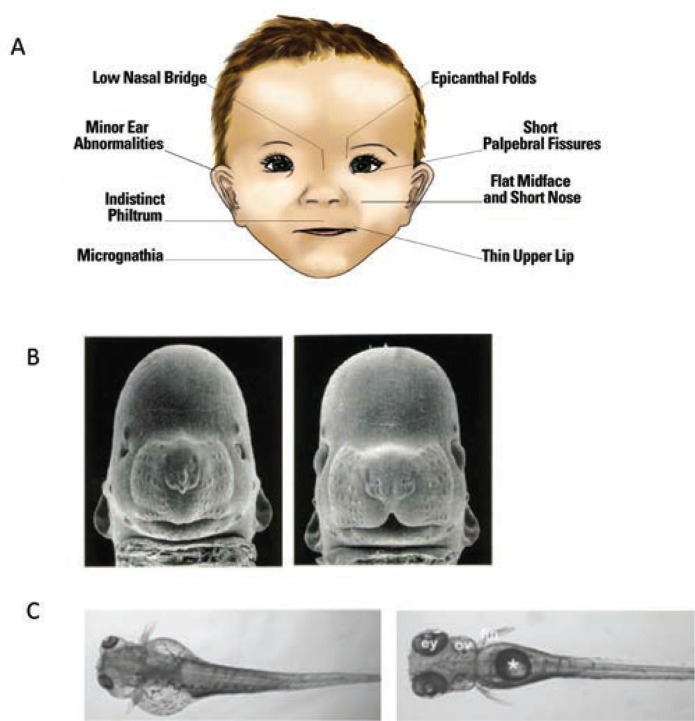
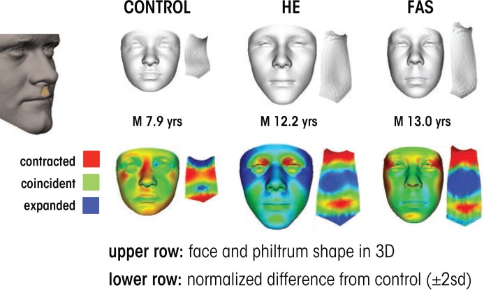
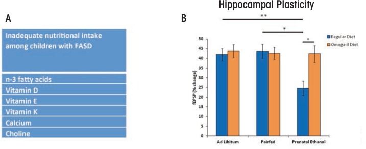
Prenatal alcohol exposure can cause a number of physical, behavioral, cognitive, and neural impairments, collectively known as fetal alcohol spectrum disorders (FASD). This article examines basic research that has been or could be translated into practical applications for the diagnosis or treatment of FASD. Diagnosing FASD continues to be a challenge, but advances are being made at both basic science and clinical levels. These include identification of biomarkers, recognition of subtle facial characteristics of exposure, and examination of the relation between face, brain, and behavior. Basic research also is pointing toward potential new interventions for FASD involving pharmacotherapies, nutritional therapies, and exercise interventions. Although researchers have assessed the majority of these treatments in animal models of FASD, a limited number of recent clinical studies exist. An assessment of this literature suggests that targeted interventions can improve some impairments resulting from developmental alcohol exposure. However, combining interventions may prove more efficacious. Ultimately, advances in basic and clinical sciences may translate to clinical care, improving both diagnosis and treatment.


Figures

Figure 1
Figure 1
Craniofacial anomalies associated with alcohol exposure during development. (A) An illustration of a child with facial features of fetal alcohol syndrome (FAS). (B) Left figure shows a mouse with gestational day 7 alcohol exposure: Note small head, small eyes, and lack of a cleft under the nose compared with the control mouse on the right. (C) Zebrafish with embryonic alcohol exposure on the left compared with a control on the right. Again notice the small eyes, the smaller head, and the malformed body cavity and fin displacement resulting from alcohol exposure. SOURCE: Figure 1A: Warren et al. 2011. Photos in B are courtesy of Dr. Kathleen Sulik, University of North Carolina at Chapel Hill. Photos in C were taken from Marrs et al. 2010.
Figure 2
Figure 2
Indirect and direct markers of alcohol exposure. (A) Ideally, biomarkers could be both sensitive and specific to alcohol exposure and also indicate the timing and amount of alcohol exposure. This figure shows the period of time, or detection window, during which alcohol consumption can be detected and the lowest levels of alcohol consumption detectable by current alcohol biomarkers. For example, fatty acid ethyl esters are detectable in a variety of biological samples, such as neonatal hair and meconium, for several months after exposure. (B) MicroRNAs (miRNAs) may serve as potential biomarkers. Using a sheep model, Dr. Rajesh Miranda has identified several miRNAs that are modified by ethanol. As shown in this panel, miR-9 expression was significantly increased in plasma from the ethanol-exposed pregnant female compared with the control female but significantly decreased in plasma from neonatal lamb compared with controls. Alterations in miR-9 may be indicative of alcohol exposure in the mother, but also may serve as a marker of alcohol-induced injury in the neonate. SOURCE: Figure 2(A): Bakhireva and Savage 2011. Figure 2(B): Modified from Balaraman et al. 2014. NOTE: * = significantly different from control.
Figure 3
Figure 3
Three-dimensional facial imaging used to detect the effects of prenatal alcohol exposure. Each case shows face and philtrum (ridge under nose) shape as well as heat maps indicating significant regions of difference from age- and sex-matched control subjects. The control case shows an unexposed individual with some flattening across the nasal bridge, a small jaw and a strongly grooved philtrum. The heavily exposed (HE) case is an individual with known exposure without clinically recognized fetal alcohol syndrome (FAS). The overall face size is average or larger and the upper part of philtrum is smooth. The FAS case shows a reduced face size and philtrum smoothness, best revealed in the philtrum heat map; red at outer canthi (outer edge of eye) identifies narrow palpebral fissures.
Figure 4
Figure 4
Magnetic resonance imaging (MRI) images showing the differential effect of different timing of exposure on face shape and brain morphology. (A) The left panel shows a control, whereas the two other panels show animals exposed on gestation day 7 and gestation day 8.5. The different timing produces differential effects on face and brain. (B) An illustration of how the shape analysis shown in figure 3 can be applied to the mouse images. The left panel shows the difference between an animal exposed on gestation day 7 versus a control. Red areas indicate a reduction in size. The middle panel shows gestation day 8 exposure versus control, note the absence of many red areas. The right panel shows the difference between the two exposure times. (C) Ethanol interacts synergistically with the PDGFRA gene. The two left most figures show an intact embryo and the dissected neurocranium of a stained PDGFRA heterozygote displaying normal morphology of the neurocranium. The right most panel shows how ethanol severely disrupts development of the anterior neurocranium and palate of the zebrafish. The homozygote, −/−, (not shown) is even more affected. SOURCE: Photos in A and B are courtesy of Dr. Kathleen Sulik, University of North Carolina at Chapel Hill. Photos in C are courtesy of Dr. Johann Eberhart, University of Texas at Austin.
Figure 5
Figure 5
(A) Many children with fetal alcohol spectrum disorder (FASD) are not consuming adequate or recommended levels of nutrients (Fuglestad et al. 2013). (B) Rodent models have shown that postnatal supplementation with various nutrients, including vitamin D, choline, and omega-3 fatty acids can reduce the severity of FASD. As shown in B, prenatal alcohol exposure in a rodent model impaired hippocampal plasticity, as measured by reduced long-term potentiation (blue bars = normal diet), an effect attenuated with postnatal supplementation with omega-3 fatty acids (orange bars = omega-3 supplemented diet) (Patten, et al. 2013b). Such studies illustrate how preclinical and clinical studies may inform one another in the development of effective interventions for FASD. NOTE: * = significant group differences at p ≤ 0.05; ** = significant group differences at p ≤ 0.01)

Similar articles

Cited by

References

    1. Avalos LA, Kaskutas L, Block G, et al. Does lack of multinutrient supplementation during early pregnancy increase vulnerability to alcohol-related preterm or small-for-gestational-age births? Maternal and Child Health Journal. 2011;15(8):1324–1332. - PMC - PubMed
    1. Bakhireva LN, Savage DD. Focus on: Biomarkers of fetal alcohol exposure and fetal alcohol effects. Alcohol Research & Health. 2011;34(1):56–63. - PMC - PubMed
    1. Balaraman S, Lunde ER, Sawant O, et al. Maternal and neonatal plasma microRNA biomarkers for fetal alcohol exposure in an ovine model. Alcoholism: Clinical and Experimental Research. 2014;38(5):1390–1400. - PMC - PubMed
    1. Bekdash RA, Zhang C, Sarkar DK. Gestational choline supplementation normalized fetal alcohol-induced alterations in histone modifications, DNA methylation, and proopiomelanocortin (POMC) gene expression in β-endorphin-producing POMC neurons of the hypothalamus. Alcoholism: Clinical and Experimental Research. 2013;37(7):1133–1142. - PMC - PubMed
    1. Bertrand J, Floyd LL, Weber MK. Guidelines for identifying and referring persons with fetal alcohol syndrome. Morbidity and Mortality Weekly Report (MMWR) Recommendations and Reports. 2005;54(RR–11):1–14. - PubMed

Publication types

MeSH terms