. 2015 Sep 8;6(26):22758-66.
doi: 10.18632/oncotarget.4283.

β-catenin stabilization enhances SS18-SSX2-driven synovial sarcomagenesis and blocks the mesenchymal to epithelial transition

Affiliations

β-catenin stabilization enhances SS18-SSX2-driven synovial sarcomagenesis and blocks the mesenchymal to epithelial transition

Jared J Barrott et al. Oncotarget. .

Abstract

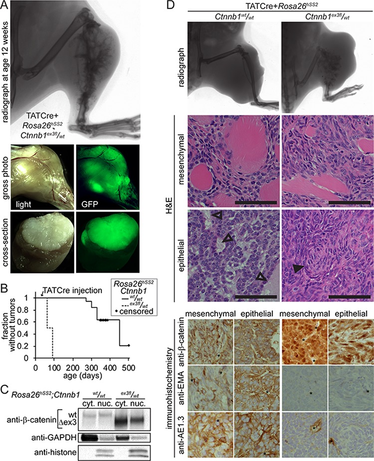
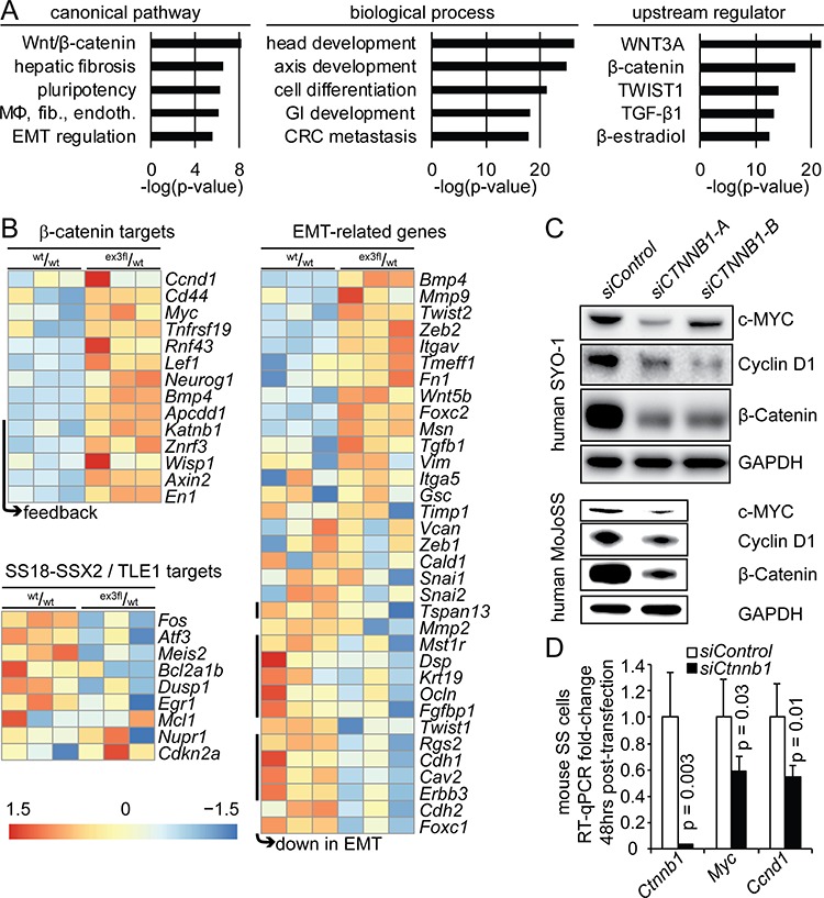
β-catenin is a master regulator in the cellular biology of development and neoplasia. Its dysregulation is implicated as a driver of colorectal carcinogenesis and the epithelial-mesenchymal transition in other cancers. Nuclear β-catenin staining is a poor prognostic sign in synovial sarcoma, the most common soft-tissue sarcoma in adolescents and young adults. We show through genetic experiments in a mouse model that expression of a stabilized form of β-catenin greatly enhances synovial sarcomagenesis. Stabilization of β-catenin enables a stem-cell phenotype in synovial sarcoma cells, specifically blocking epithelial differentiation and driving invasion. β-catenin achieves its reprogramming in part by upregulating transcription of TCF/LEF target genes. Even though synovial sarcoma is primarily a mesenchymal neoplasm, its progression towards a more aggressive and invasive phenotype parallels the epithelial-mesenchymal transition observed in epithelial cancers, where β-catenin's transcriptional contribution includes blocking epithelial differentiation.

Keywords: Wnt-signaling; epithelial-mesenchymal transition; mouse genetic model; translocation.


Conflict of interest statement

CONFLICTS OF INTEREST

The authors declare no conflicts of interest.

Figures

Figure 1
Figure 1. β-catenin stabilization enhances SS18-SSX2-driven tumorigenesis
A. Schematics represent the conditional alleles for expression of SS18-SSX2 from the Rosa26 locus, Luciferase from a CAG promoter in the 3′ untranslated region of the RNA polymerase 2 locus, and removal of the third exon in Ctnnb1. Total RNA and protein obtained from mouse embryonic fibroblasts and analyzed by RT-PCR and Western demonstrate relative stabilization at the protein level of the β-catenin isoform resulting from removal of the third exon (Δex3). B. Tabulated tumor formation in 3 groups of mice bearing Rosa26hSS2 and/or Ctnnb1ex3fl, injected at 4 weeks with AdCre. Radiographs and luciferase luminescence overlay images of these tumors 12 weeks after AdCre injection. C. Localized injection of AdCre into the pretibial soft-tissues leads to early tumor development outside the bone and reactive osteoid formation detectable at 2 and 4 weeks after the injection. (Arrows indicate the normal position of periosteum, here replaced by tumor in each example.) D. Representative histologic sections from the same cohort of mice stained with H&E demonstrate the classic SS histopathology, including hemangiopericytomatous vascular spaces (black arrow) and poorly differentiated areas with cells dominated by large nuclei and scant cytoplasm. Immunohistochemical stains demonstrate abundant nuclear TLE1 and cytoplasmic BCL2 in these tumors. (Magnification bars are 40 μm. Immuno-photomicrograph panels are 40 μm square.)
Figure 2
Figure 2. Tumors developing in mice with stabilized β-catenin are synovial sarcomas by transcriptome
A. Heat map of a list of genes previously determined to be enriched in human and mouse SSs, comparing control skeletal muscle (noted by m) to two mouse models of SS without conditional genetic stabilization of Ctnnb1 (noted by triangles) and the current model wherein β-catenin is stabilized concurrent to expression of SS18-SSX2, induced by AdCre injection into the hind limb soft-tissues (noted by squares). B. Unsupervised hierarchical clustering of these three models of mouse SS, compared to 78 other genetically-induced mouse tumor models.
Figure 3
Figure 3. Initiation of tumors with injected TATCre enables comparison of SS18-SSX2-induced tumors with and with genetic stabilization of β-catenin
A. Radiograph, gross and cross sectional photographs each with light and GFP fluorescence demonstrate the typical tumor that has developed by 8 weeks after TATCre injection into Rosa26hSS2/wt; Ctnnb1ex3fl/wt mice at age 4 weeks. B. Kaplan-Meier plot of the relative latency to tumorigenesis following TATCre injection at age 4 weeks into Rosa26hSS2/wt mice with (n = 16) or without (n = 19) an ex3f l allele of Ctnnb1. C. Westerns of cytoplasmic (cyt.) and nuclear (nuc.) protein fractions from tumors demonstrate degradation to below detectable levels of full length β-catenin in tumors heterozygous for the stabilized, Δex3 form, suggesting feedback activation. D. Comparison of radiographic evidence of skeletal element invasion, muscle invasion by monophasic areas of tumor cells, the maximal epithelial differentiation, and immunophenotype of both monophasic and biphasic areas of TATCre-induced Rosa26hSS2/wt tumors with or without stabilized β-catenin. (Magnification bars are 50 μm. Immunohistochemistry photomicrographs are 50 μm-square panels. Open arrows indicate epithelial glands. Filled arrows indicate swirls of mesenchymal appearing cells, the structure most similar to glands that formed in tumors with stabilized β-catenin. * indicates position of vessel.)
Figure 4
Figure 4. Stabilization of β-catenin drives an EMT transcriptional signature and increases expression of TCF/LEF target genes
A. Chart of the log of the p-values for the top 5 canonical pathways, biological processes, and upstream regulators from ingenuity pathway analysis on the genes that were differentially expressed between TATCre-initiated Rosa26hSS2 tumors with and without stabilized β-catenin. (MΦ, fib., endoth. indicates the macrophages, fibroblasts and endothelial cells activated in rheumatoid arthritis. GI indicates gastrointestinal. CRC indicates colorectal carcinoma.) B. Heat maps of differentially expressed β-catenin target genes, target genes of SS18-SSX2-mediated transcriptional repression via TLE1, and genes related to the epithelial-to-mesenchymal transition (EMT). C. Western blots from two human SS cell lines 72 hours after transfection with either control or anti-CTNNB1 small interfering RNA (siRNA) molecules. D. Chart of fold-expression by RT-qPCR of Myc and Ccnd1 48 hours following siRNA against Ctnnb1 in mouse synovial sarcoma cells.

References

    1. Herzog CE. Overview of sarcomas in the adolescent and young adult population. J Pediatr Hematol Oncol. 2005;27:215–8. - PubMed
    1. Ladanyi M, Antonescu CR, Leung DH, Woodruff JM, Kawai A, Healey JH, Brennan MF, Bridge JA, Neff JR, Barr FG, Goldsmith JD, Brooks JS, Goldblum JR, Ali SZ, Shipley J, Cooper CS, Fisher C, Skytting B, Larsson O. Impact of SYT-SSX fusion type on the clinical behavior of synovial sarcoma: a multi-institutional retrospective study of 243 patients. Cancer Res. 2002;62:135–40. - PubMed
    1. Haldar M, Hancock JD, Coffin CM, Lessnick SL, Capecchi MR. A conditional mouse model of synovial sarcoma: insights into a myogenic origin. Cancer cell. 2007;11:375–88. - PubMed
    1. Moon RT, Kohn AD, De Ferrari GV, Kaykas A. WNT and beta-catenin signalling: diseases and therapies. Nat Rev Genet. 2004;5:691–701. - PubMed
    1. Liu C, Li Y, Semenov M, Han C, Baeg GH, Tan Y, Zhang Z, Lin X, He X. Control of beta-catenin phosphorylation/degradation by a dual-kinase mechanism. Cell. 2002;108:837–47. - PubMed

Publication types

LinkOut - more resources