. 2015 Sep 15;195(6):2591-600.
doi: 10.4049/jimmunol.1501138. Epub 2015 Aug 10.

IL-17A Contributes to the Pathogenesis of Endometriosis by Triggering Proinflammatory Cytokines and Angiogenic Growth Factors

Affiliations

IL-17A Contributes to the Pathogenesis of Endometriosis by Triggering Proinflammatory Cytokines and Angiogenic Growth Factors

Soo Hyun Ahn et al. J Immunol. .

Abstract

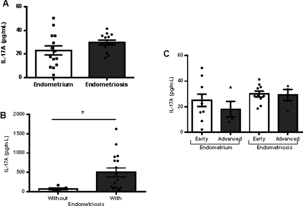
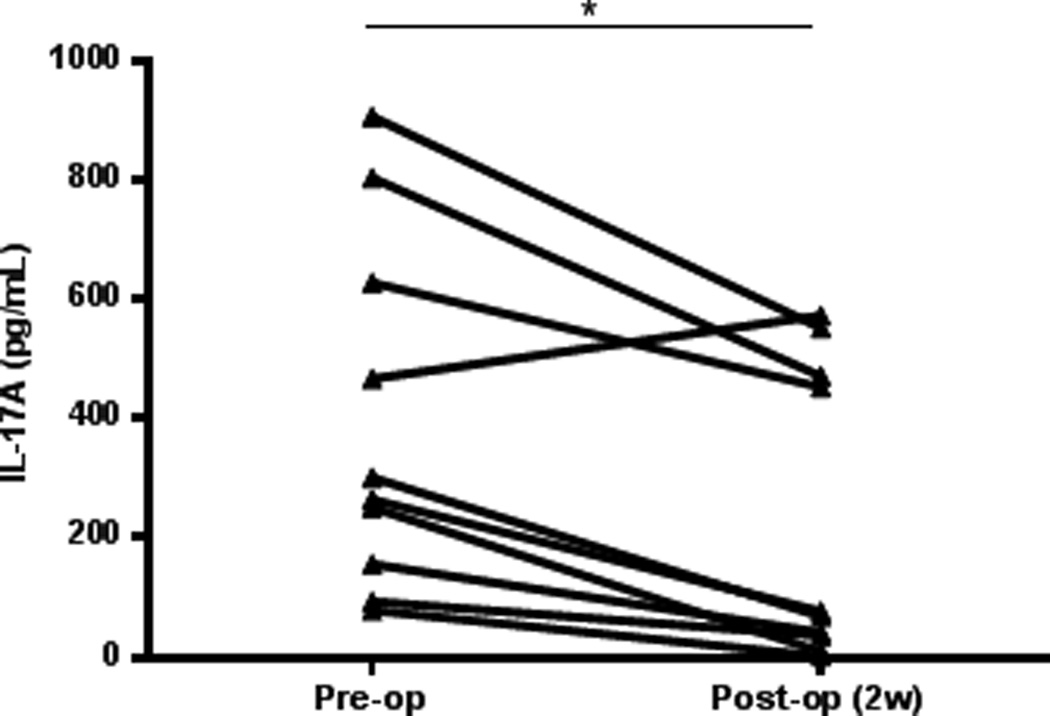
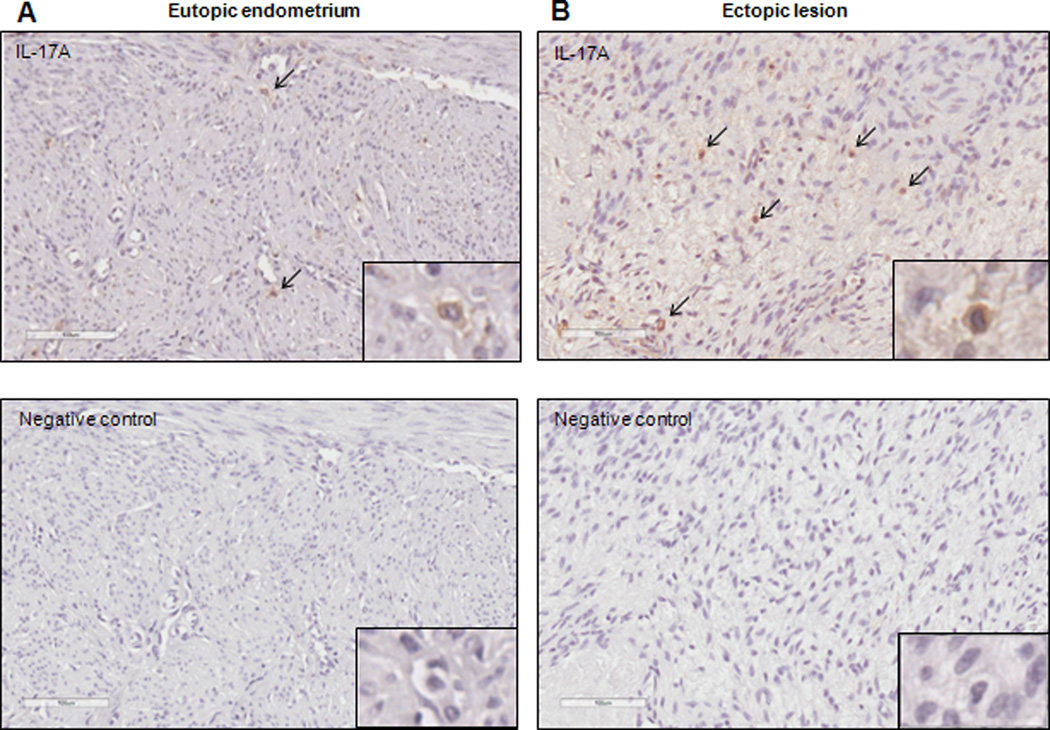
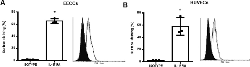
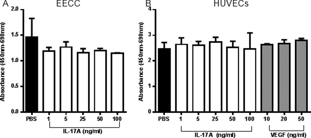
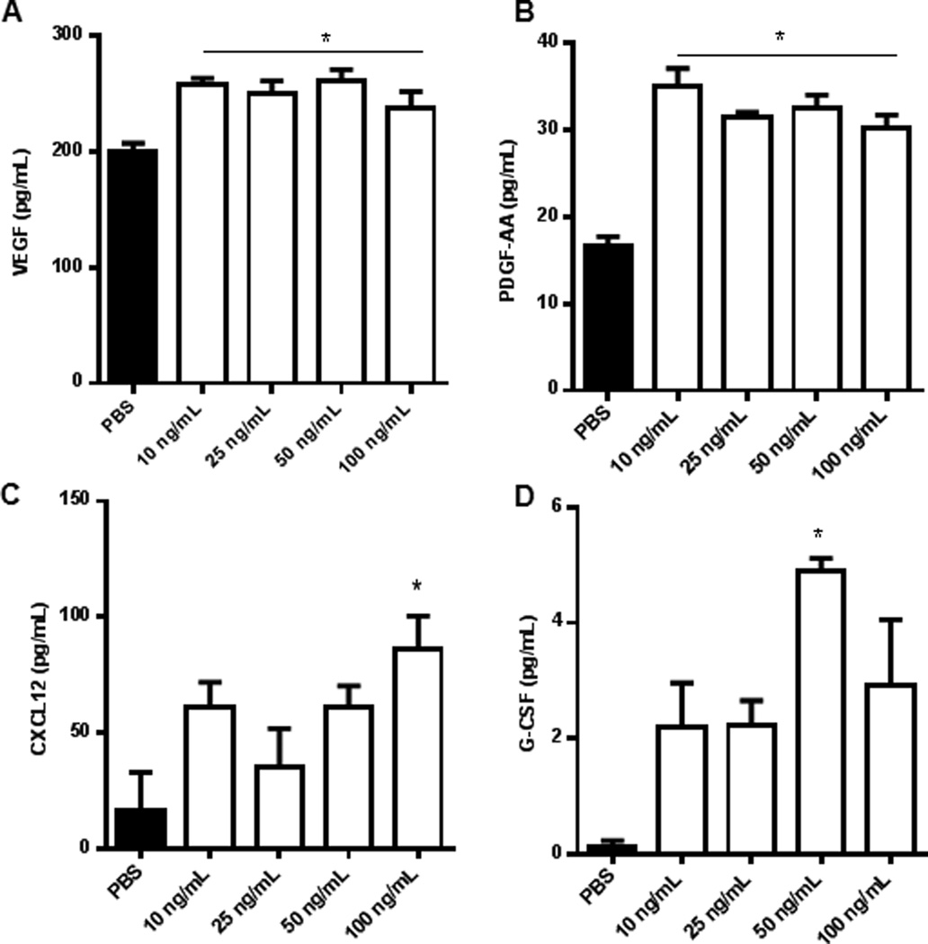
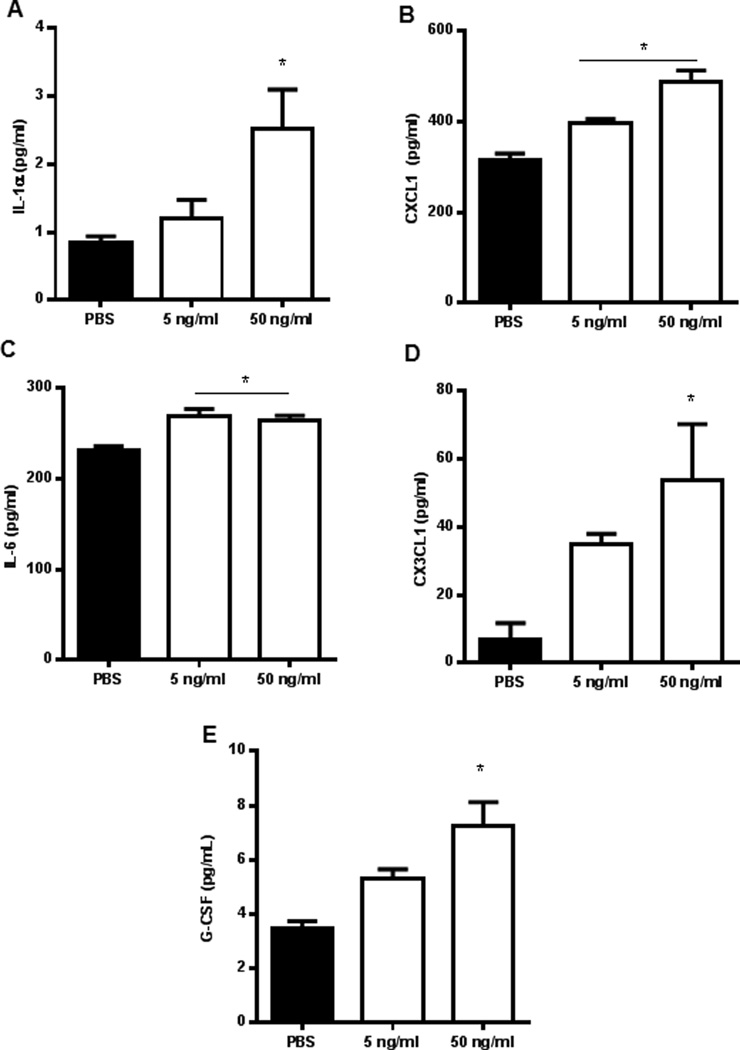
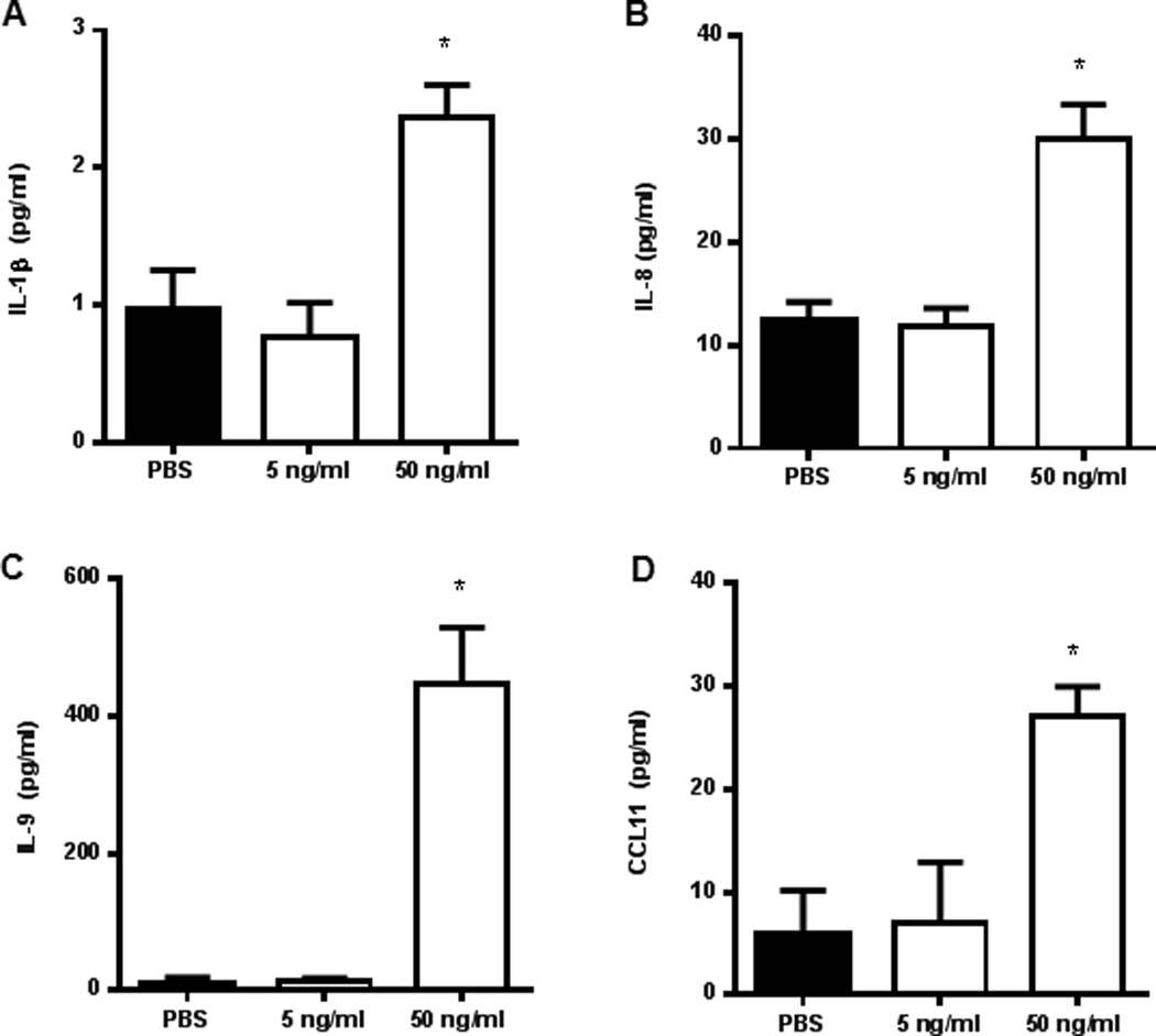
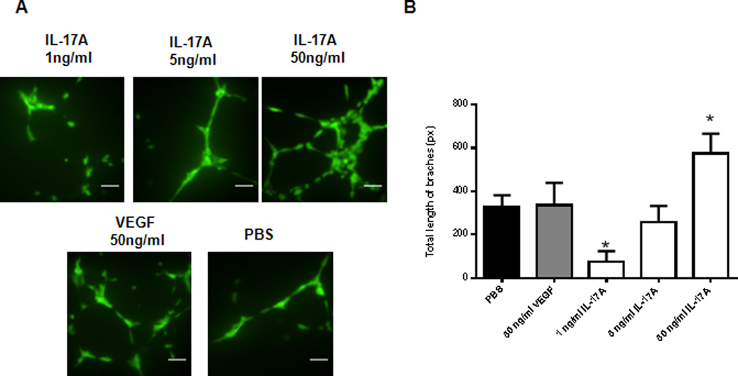
Endometriosis is a chronic, inflammatory disease characterized by the growth of endometrial tissue in aberrant locations outside the uterus. Neoangiogenesis or establishment of new blood supply is one of the fundamental requirements of endometriotic lesion survival in the peritoneal cavity. IL-17A is emerging as a potent angiogenic and proinflammatory cytokine involved in the pathophysiology of several chronic inflammatory diseases such as rheumatoid arthritis and psoriasis. However, sparse information is available in the context of endometriosis. In this study, we demonstrate the potential importance of IL-17A in the pathogenesis and pathophysiology of endometriosis. The data show a differential expression of IL-17A in human ectopic endometriotic lesions and matched eutopic endometrium from women with endometriosis. Importantly, surgical removal of lesions resulted in significantly reduced plasma IL-17A concentrations. Immunohistochemistry revealed localization of IL-17A primarily in the stroma of matched ectopic and eutopic tissue samples. In vitro stimulation of endometrial epithelial carcinoma cells, Ishikawa cells, and HUVECs with IL-17A revealed significant increase in angiogenic (vascular endothelial growth factor and IL-8), proinflammatory (IL-6 and IL-1β), and chemotactic cytokines (G-CSF, CXCL12, CXCL1, and CX3CL1). Furthermore, IL-17A promoted tubulogenesis of HUVECs plated on Matrigel in a dose-dependent manner. Thus, we provide the first evidence, to our knowledge, that endometriotic lesions produce IL-17A and that the removal of the lesion via laparoscopic surgery leads to the significant reduction in the systemic levels of IL-17A. Taken together, our data show a likely important role of IL-17A in promoting angiogenesis and proinflammatory environment in the peritoneal cavity for the establishment and maintenance of endometriosis lesions.


Figures

Figure 1
Figure 1
IL-17A expression in tissue and plasma samples. (A) Matched eutopic endometrium (n=14, 23.0±3.84pg/ml) and ectopic lesions (n=14, 29.77±1.97pg/ml) (B) Plasma samples from women without (n=7, 72.90±18.94pg/ml) and with (n=17, 499.4±116.4pg/ml) endometriosis. IL-17A levels were significantly higher in women with endometriosis compared to healthy controls (*p<0.05). (C) IL-17A concentration in matched eutopic endometrium and ectopic lesions distributed by disease severity following ASRM staging criteria (early denotes Stages I-II, and advanced denotes Stages III-IV). All data are represented as mean ± SEM.
Figure 2
Figure 2
IL-17A concentration in plasma of women (n=10) undergoing laparoscopy surgery for the removal of endometriosis. IL-17A concentration was measured from the patient prior to undergoing surgery and 2 weeks post-surgical visit to the clinic. The concentration of IL-17A diminishes significantly in the peripheral blood of women with endometriosis after lesion is removed (*p=0.0016).
Figure 3
Figure 3
IL-17A positive cells are detected in the matched eutopic (A) and ectopic lesion (B) samples from women with endometriosis. Immunohistochemistry images are representative of 5 matched tissue samples immunostained with anti-human IL-17A. 200× magnification with digitally magnified inlet; scale bar represents 100µm.
Figure 4
Figure 4
IL-17RA is expressed on EECCs and HUVECs. EECCs (A) and HUVECs (B) were stained with either PE conjugated mouse anti-human IL-17RA or PE conjugated Mouse IgG isotype control in room temperature. On average 65.2±3.9% of EECCs stained for IL-17RA whereas 58.1±14.6% cell surface staining was seen for HUVECs. Representative of three separate experiments. *p<0.05 compared to isotype.
Figure 5
Figure 5
WST-1 proliferation assay indicate that IL-17A does not induce proliferation of EECCs and HUVECs in vitro. (A) EECCs were treated with different concentrations of IL-17A (1, 5, 25, 50 and 100 ng/ml) or PBS control to assess the effect of IL-17A on proliferation. (B) HUVECs were treated with different concentrations of IL-17A (1, 5, 25, 50 and 100 ng/ml) or PBS control to assess the effect of IL-17A on proliferation. VEGF (10, 20, 50 ng/ml) was used as a positive control. Three separate WST-1 proliferation assay were conducted on both (A) and (B) as per standard protocol.
Figure 6
Figure 6
IL-17A induces production of chemokine and angiogenic cytokines from EECCs. Endometrial epithelial carcinoma cells (EECCs) were plated onto a 96-well cell culture plate in triplicate at a density of 5×10⁵cells/well and incubated with different concentrations of IL-17A (10, 25, 50, 100ng/mL) for 24 hours at 37°C with 5% CO2. The conditioned supernatants of EECCs were collected and screened for cytokine expression from which PDGF-AA, VEGF, CXCL12 and G-CSF showed statistical significance. *p < 0.05 compared with PBS.
Figure 7
Figure 7
IL-17A induces the production of pro-inflammatory cytokines and chemokines from HUVECs in a dose dependent manner. HUVECs were plated onto a 6-well plate in triplicate at a density of 1×10⁵ cells/plate and were incubated with different concentrations of IL-17A (5 and 50 ng/ml) for 24 hours in a standard cell culture incubator at 37°C with 5% CO2. Conditioned supernatants were collected and screened for cytokine expression from which IL-1α, CXCL1, IL-6, CX3CL1, and G-CSF (A-E, respectively) showed statistical significance. *p<0.05 compared with PBS.
Figure 8
Figure 8
IL-17A induces production of chemokine, angiogenic and pro-inflammatory cytokines from Ishikawa cells. Ishikawa cells were plated onto a 96-well cell culture plate in triplicate at a density of 5×10⁵cells/well and incubated with different concentrations of IL-17A (5 and 50 ng/mL) or PBS control for 24 hours at 37°C with 5% CO2. The conditioned supernatants of each treatment were collected and screened for cytokine expression from which IL-1β, IL-8, IL-9 and CCL11 showed statistical significance. *p < 0.05 compared with PBS.
Figure 9
Figure 9
IL-17A promotes in vitro tubulogenesis in HUVECs. (A) HUVECs were plated on Matrigel at 5×10³ cells/well using Ibidi µ-slide angiogenesis plate (Cat. #81506) in triplicate per treatment. VEGF (50ng/ml) and PBS were used as a positive and negative control, respectively. Scale bar represents 10µm. 150× magnification using a confocal microscope (Quorum Wave Effects Spinning Disc Confocal, Queen’s University Cancer Research Institute Imaging Facility) (B) Total length of branches in the field of image was measured using ImageJ Angiogenesis Analyzer Macro with HUVECs phase contrast setting. *p<0.05 compared with PBS.

References

    1. Murphy AA. Clinical Aspects of Endometriosis. Ann N Y Acad Sci. 2002;955:1–10. - PubMed
    1. Vercellini P, Vigano P, Somigliana E, Fedele L. Endometriosis: pathogenesis and treatment. Nat Rev Endocrinol. 2014;10:261–275. - PubMed
    1. Edwards AK, Nakamura DS, Virani S, Wessels JM, Tayade C. Animal models for anti-angiogenic therapy in endometriosis. J Reprod Immunol. 2013;97:85–94. - PubMed
    1. Laschke MW, Menger MD. Anti-angiogenic treatment strategies for the therapy of endometriosis. Hum Reprod Update. 2012;18:682–702. - PubMed
    1. Nap AW, Griffoen AW, Dunselman GA, Bouma-Ter Steege JC, Thijssen VL, Evers JL, Groothuis PG. Antiangiogenic therapy for endometriosis. J Clin Endocrinol Metab. 2004;89:1089–1095. - PubMed

Publication types

MeSH terms