

Dynamic markers based on blood perfusion fluctuations for selecting skin melanocytic lesions for biopsy
- PMID: 26260030
- PMCID: PMC4542467
- DOI: 10.1038/srep12825
Dynamic markers based on blood perfusion fluctuations for selecting skin melanocytic lesions for biopsy
Erratum in
-
Erratum: Dynamic markers based on blood perfusion fluctuations for selecting skin melanocytic lesions for biopsy.Sci Rep. 2016 Feb 9;6:20162. doi: 10.1038/srep20162. Sci Rep. 2016. PMID: 26859727 Free PMC article. No abstract available.
Abstract
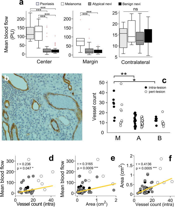
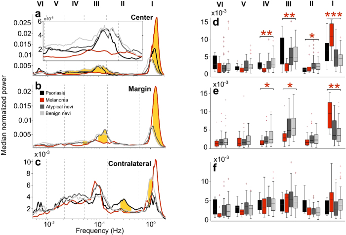
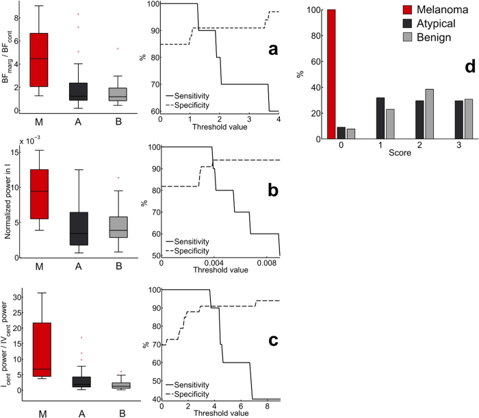
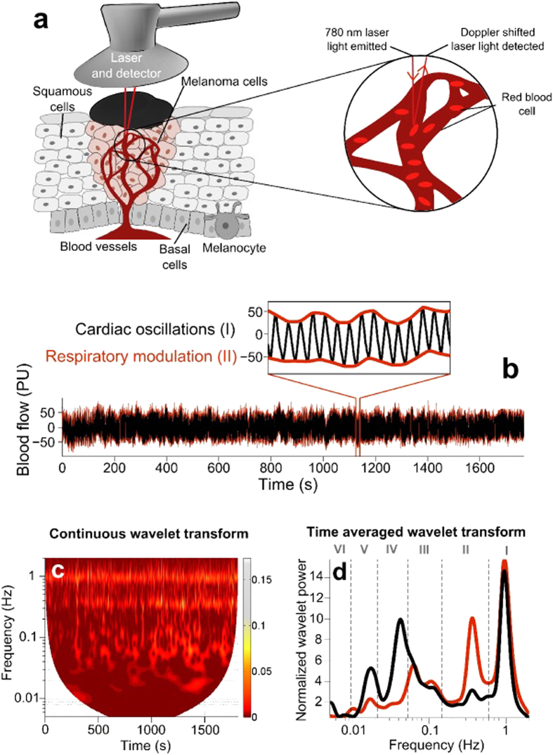
Skin malignant melanoma is a highly angiogenic cancer, necessitating early diagnosis for positive prognosis. The current diagnostic standard of biopsy and histological examination inevitably leads to many unnecessary invasive excisions. Here, we propose a non-invasive method of identification of melanoma based on blood flow dynamics. We consider a wide frequency range from 0.005-2 Hz associated with both local vascular regulation and effects of cardiac pulsation. Combining uniquely the power of oscillations associated with individual physiological processes we obtain a marker which distinguishes between melanoma and atypical nevi with sensitivity of 100% and specificity of 90.9%. The method reveals valuable functional information about the melanoma microenvironment. It also provides the means for simple, accurate, in vivo distinction between malignant melanoma and atypical nevi, and may lead to a substantial reduction in the number of biopsies currently undertaken.
Figures
Similar articles
-
Development of a novel noninvasive adhesive patch test for the evaluation of pigmented lesions of the skin.J Am Acad Dermatol. 2014 Aug;71(2):237-44. doi: 10.1016/j.jaad.2014.04.042. Epub 2014 Jun 4. J Am Acad Dermatol. 2014. PMID: 24906614
-
Comparison of pHH3, Ki-67, and survivin immunoreactivity in benign and malignant melanocytic lesions.Am J Dermatopathol. 2008 Apr;30(2):117-22. doi: 10.1097/DAD.0b013e3181624054. Am J Dermatopathol. 2008. PMID: 18360113
-
Follow-up of melanocytic skin lesions with digital epiluminescence microscopy: patterns of modifications observed in early melanoma, atypical nevi, and common nevi.J Am Acad Dermatol. 2000 Sep;43(3):467-76. doi: 10.1067/mjd.2000.107504. J Am Acad Dermatol. 2000. PMID: 10954658
-
Distinguishing melanocytic nevi from melanoma by DNA copy number changes: comparative genomic hybridization as a research and diagnostic tool.Dermatol Ther. 2006 Jan-Feb;19(1):40-9. doi: 10.1111/j.1529-8019.2005.00055.x. Dermatol Ther. 2006. PMID: 16405569 Review.
-
Dermoscopy patterns of nevi associated with melanoma.G Ital Dermatol Venereol. 2010 Feb;145(1):99-110. G Ital Dermatol Venereol. 2010. PMID: 20197749 Review.
Cited by
-
Spatial analysis of photoplethysmography in cutaneous squamous cell carcinoma.Sci Rep. 2022 May 5;12(1):7318. doi: 10.1038/s41598-022-10924-3. Sci Rep. 2022. PMID: 35513459 Free PMC article.
-
In-vivo correlations between skin metabolic oscillations and vasomotion in wild-type mice and in a model of oxidative stress.Sci Rep. 2019 Jan 17;9(1):186. doi: 10.1038/s41598-018-36970-4. Sci Rep. 2019. PMID: 30655574 Free PMC article.
-
Dermatology Roundup: The Latest Tips, Techniques, and Technologies for Busy Clinicians.J Clin Aesthet Dermatol. 2017 Mar;10(3):S26-S31. Epub 2017 Mar 1. J Clin Aesthet Dermatol. 2017. PMID: 28360972 Free PMC article. Review.
-
Bifurcation in Blood Oscillatory Rhythms for Patients with Ischemic Stroke: A Small Scale Clinical Trial using Laser Doppler Flowmetry and Computational Modeling of Vasomotion.Front Physiol. 2017 Mar 23;8:160. doi: 10.3389/fphys.2017.00160. eCollection 2017. Front Physiol. 2017. PMID: 28386231 Free PMC article.
-
Comparison of pancreatic microcirculation profiles in spontaneously hypertensive rats and Wistar-kyoto rats by laser doppler and wavelet transform analysis.Physiol Res. 2020 Dec 22;69(6):1039-1049. doi: 10.33549/physiolres.934448. Epub 2020 Nov 2. Physiol Res. 2020. PMID: 33129246 Free PMC article.
References
-
- American Cancer Society. Cancer Facts & Figures 2013. http://www.cancer.org/research/cancerfactsfigures/cancer-facts-figures-2013. Date of access: 24/03/2014.
-
- Vestergaard M. E., Macaskill P., Holt P. E. & Menzies S. W. Dermoscopy compared with naked eye examination for the diagnosis of primary melanoma: a meta-analysis of studies performed in a clinical setting. Br. J. Dermatol. 159, 669–676 (2008). - PubMed
-
- Argenziano G., Soyer H. P., Chimenti S. & Ruocco V. Impact of dermoscopy on the clinical management of pigmented skin lesions. Clin. Dermatol. 20, 200–202 (2002). - PubMed
-
- Kittler H., Pehamberger H., Wolff K. & Binder M. Diagnostic accuracy of dermoscopy. Lancet Oncol. 3, 159–165 (2002). - PubMed
-
- Patel J. K. et al. Newer technologies/techniques and tools in the diagnosis of melanoma. Eur. J. Dermatol. 18, 617–631 (2008). - PubMed
Publication types
MeSH terms
Substances
LinkOut - more resources
Full Text Sources
Other Literature Sources
Medical
