. 2015 Aug 11:14:152.
doi: 10.1186/s12943-015-0417-y.

A critical role of Oct4A in mediating metastasis and disease-free survival in a mouse model of ovarian cancer

Affiliations

A critical role of Oct4A in mediating metastasis and disease-free survival in a mouse model of ovarian cancer

Chantel Samardzija et al. Mol Cancer. .

Abstract

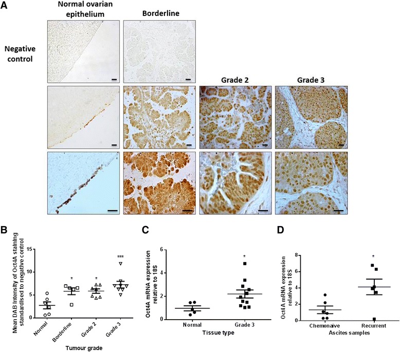
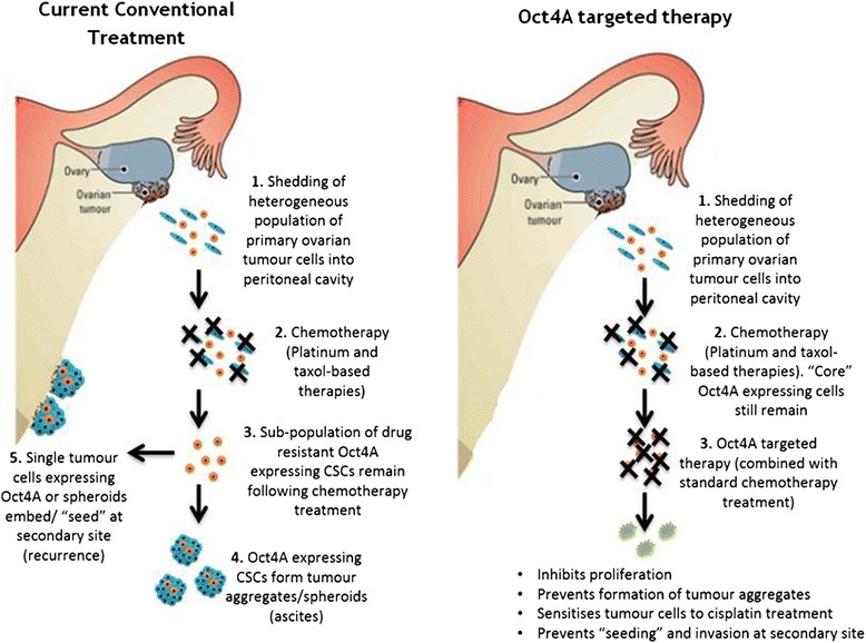
Background: High grade epithelial ovarian cancer (EOC) is commonly characterised by widespread peritoneal dissemination and ascites. Metastatic EOC tumour cells can attach directly to neighbouring organs or alternatively, maintain long term tumourigenicity and chemoresistance by forming cellular aggregates (spheroids). Cancer stem-like cells are proposed to facilitate this mechanism. This study aimed to investigate the role of Oct4A, an embryonic stem cell factor and known master regulator of pluripotency in EOC progression, metastasis and chemoresistance.

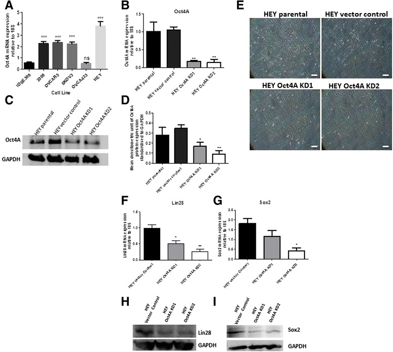
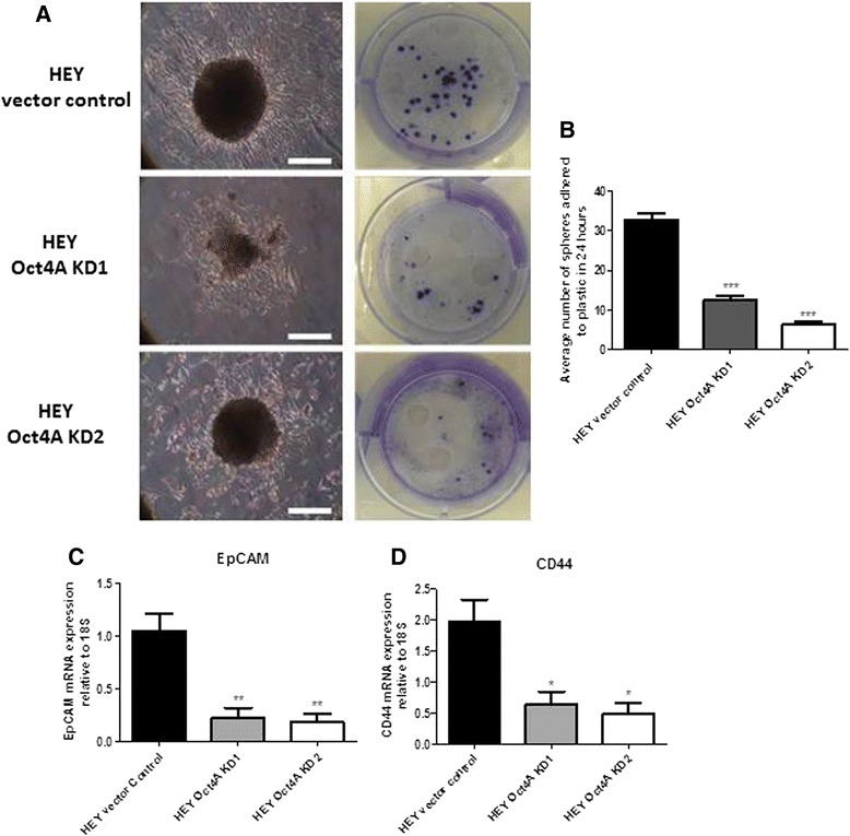
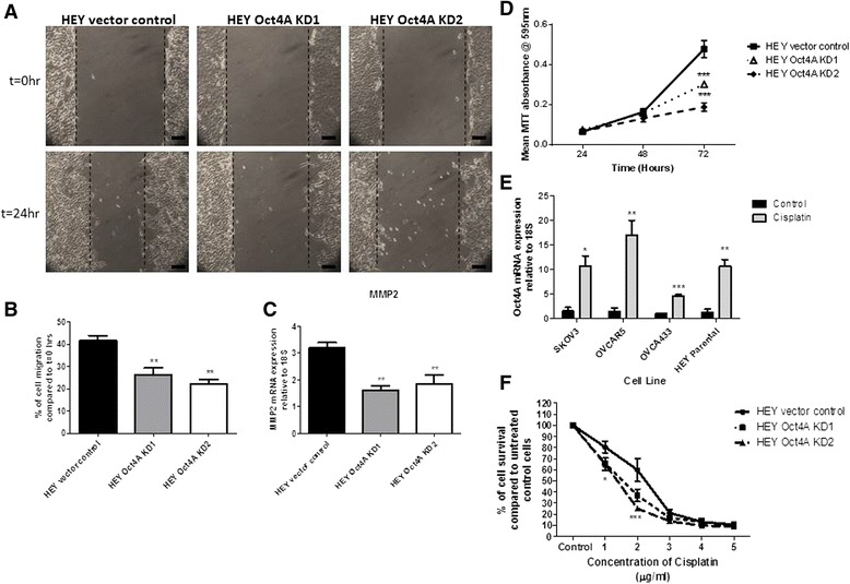
Methods: To investigate the expression of Oct4A in primary EOC tumours, IHC and qRT-PCR analyses were used. The expression of Oct4A in chemonaive and recurrent EOC patient ascites-derived tumour cells samples was investigated by qRT-PCR. The functional role of Oct4A in EOC was evaluated by generating stable knockdown Oct4A clones in the established EOC cell line HEY using shRNA-mediated silencing technology. Cellular proliferation, spheroid forming ability, migration and chemosensitivty following loss of Oct4A in HEY cells was measured by in vitro functional assays. These observations were further validated in an in vivo mouse model using intraperitoneal (IP) injection of established Oct4A KD clones into Balb/c nu/nu mice.

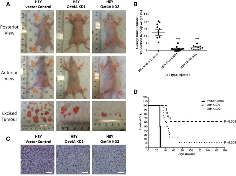
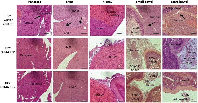
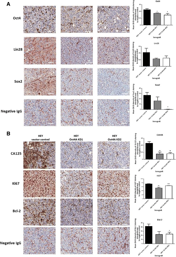
Results: We demonstrate that, compared to normal ovaries Oct4A expression significantly increases with tumour dedifferentiation. Oct4A expression was also significantly high in the ascites-derived tumour cells of recurrent EOC patients compared to chemonaive patients. Silencing of Oct4A in HEY cells resulted in decreased cellular proliferation, migration, spheroid formation and increased chemosensitivity to cisplatin in vitro. IP injection of Oct4A knockdown cells in vivo produced significantly reduced tumour burden, tumour size and invasiveness in mice, which overall resulted in significantly increased mouse survival rates compared to mice injected with control cells.

Conclusions: This data highlights a crucial role for Oct4A in the progression and metastasis of EOC. Targeting Oct4A may prove to be an effective strategy in the treatment and management of epithelial ovarian tumours.


Figures

Fig. 1
Fig. 1
Expression and localization of Oct4A in primary serous epithelial ovarian tumours. a Representative immunohistochemical staining of Oct4A in normal (n = 7), borderline (n = 5), grade 2 (n = 7) and grade 3 (n = 8) primary ovarian tumours. Positive Oct4A expression is indicated by intense nuclear staining. Images are set at 200x (top panels including negative controls) and 400x (bottom panels). Scale bars represent 10 μM. b Quantification of Oct4A staining using Image J software which recognizes total DAB intensity. Variations in staining were determined by subtracting the negative control DAB reading from the Oct4A DAB reading for each tissue sample. Data is presented as the mean ± SEM of Oct4A DAB staining intensity. Significance is indicated by *P < 0.05, **P < 0.01 or ***P < 0.001 as determined by One-way ANOVA using Dunnett’s Multiple Comparison post-test compared to normal ovarian epithelium tissue. c Oct4A mRNA expression in primary normal ovarian epithelium tissues (n = 6) and primary Grade 3 serous epithelial ovarian tumours (n = 11) was determined by qPCR analysis. Relative quantification of Oct4A mRNA expression is standardized to 18S housekeeping gene and normalized to normal ovarian epithelium tissues. Data is expressed as the mean ± SEM of log2 transformed data of samples performed in triplicate. Significant increase of Oct4A expression in Grade 3 serous EOC tumours is indicated by *P < 0.05 as determined by student's t-test. d qPCR analysis of Oct4A expression in tumour cells isolated from the ascites of chemonaive (n = 6) and recurrent (n = 6) serous EOC patients. Relative quantification of Oct4A mRNA expression was standardized to 18S housekeeping gene. Data is expressed as the mean ± SEM of Oct4A mRNA expression of each sample performed in triplicate. Significance is indicated by *P < 0.05 as determined by student's t-test
Fig. 2
Fig. 2
Stable shRNA knockdown of Oct4A in epithelial ovarian cancer cell line HEY. a Four established epithelial ovarian cancer cell lines OVCAR5, SKOV3 OVCAR433 and HEY were screened for endogenous expression of Oct4A mRNA by qPCR analysis. Relative quantification of Oct4A mRNA expression was standardized to 18S housekeeping gene and normalized to the normal ovarian epithelium immortalized cell line ISOE398. b shRNA-mediated silencing of Oct4A in HEY cells (Oct4A KD1 and Oct4A KD2) was confirmed by qPCR analysis. Relative Oct4A levels were normalized to 18S housekeeping gene and calibrated to vector control samples prepared in triplicate. c Western blot analysis using nuclear cell lysates and 12 % SDS-PAGE gels further validating Oct4A knockdown in Oct4A KD cells as determined by protein bands present at ~50 kDa. Total protein load was determined by stripping and re-probing the membrane with GAPDH. d Oct4A protein expression was significantly reduced in Oct4A KD cells as determined by densitometry analysis. Data is expressed as a ratio of Oct4A protein expression compared to GAPDH protein expression. e Phase contrast images of exponentially growing HEY cells cultured on plastic indicating no obvious morphological changes following shRNA-mediated knockdown of Oct4A. Magnification is set at 100x. Scale bar represents 50 μM. f-g Lin28 and Sox2 mRNA expression was investigated by qPCR analysis. Relative Lin28 and Sox2 levels were normalized to 18S housekeeping gene and calibrated to vector control samples prepared in triplicate. h-i Lin28 and Sox2 protein expression in Oct4A KD cells was investigated by Western blot analysis. Cytoplasmic cell lysates were prepared for Lin28 analysis and nuclear cell lysates used for Sox2 analysis. Total protein load was determined by stripping and re-probing the membrane with GAPDH. For all graphs sets, data is expressed as the mean fold change ± SEM from three independent samples. Significance is indicated by *p < 0.05, **p < 0.01 and ***p < 0.001 and determined by One-Way ANOVA using Dunnett's post-test compared to the ISOE398 cell line or HEY vector control
Fig. 3
Fig. 3
Suppression of Oct4A resulted in reduced spheroid forming ability and a loss of Lin28 and Sox2 in HEY cells. a The spheroid forming ability of HEY cells following stable shRNA-mediated knockdown of Oct4A was assessed by culturing cells on ultra-low attachment plates as described in the Materials and Methods. Cellular aggregation was monitored over 18 days and documented using phase contrast microscopy. Images are representative of three independent experiments set at 100x magnification. Scale bar represents 100 μM. b The number of spheroids produced after 18 days was assessed using a phase contrast microscope calibrated with an ocular micrometer. Spheroids with a diameter greater than 200 μM were classified as spheroids. c Expression of Oct4A, Lin28 and Sox2 in Oct4A KD 18 day spheroids was evaluated by qPCR analysis. Relative quantification of Oct4A, Lin28 and Sox2 mRNA expression in Oct4A knockdown cells was standardized to 18S housekeeping gene and normalized to vector control cells
Fig. 4
Fig. 4
Loss of Oct4A expression resulted in reduced adhesion abilities of spheroids and colony formation in HEY cells. a Vector control and Oct4A KD cells were grown as non-adherent spheroids cultures on ultra-low attachment plates as described in the Material and Methods. Following 18 day incubation, spheroids were transferred to 6 well plastic culture plates and incubated for 24 h. Adhered spheroids were then fixed with formalin and stained with 5 % crystal violet. All images are representative of three independent experiments performed in duplicate. Magnification 100x and scale = 100 μM b Colony formation in 18 day Oct4A KD spheroids was significantly reduced when compared to vector control spheroids. c Expression of adhesion markers CD44 and EpCAM in monolayer Oct4A KD cells was determined by qPCR analysis. Relative quantification CD44 and EpCAM mRNA expression in Oct4A KD cells were standardized to 18S housekeeping gene and normalized to the vector control. All data sets are presented as the mean ± SEM of three independent experiments as determined by One-Way ANOVA and Dunnett's Multiple Comparison post-test compared to vector control. Significance is indicated by *P < 0.05, **p < 0.01 and ***P < 0.001
Fig. 5
Fig. 5
Loss of Oct4A reduced migratory and proliferative abilities in HEY cells while increasing chemosensitivity to cisplatin treatment. a The migratory ability of Oct4A KD cells was determined by 24 h wound healing assay. Cells were grown as confluent monolayer prior to the insertion of a single wound and allowed to migrate for 24 h. Images of the wounds were taken at t = 0 h and t = 24 h post wound insertion and are representative of three independent experiments. Images are at 100x magnification and scale bar represents 50 μM. b 24 h migration expressed as a percentage of wound closure compared to t = 0 h using an ocular micrometer. Data is presented as the mean ± SEM of three independent experiments. Significance is indicated by **p < 0.01 as determined by One-Way ANOVA compared to vector control. c qPCR analysis of MMP2 mRNA expression in Oct4A KD monolayer cells. Relative quantification of MMP2 mRNA expression in Oct4A knockdown cells was standardized to 18S housekeeping gene and normalized the vector control. d Proliferative potential of Oct4A KD cells was determined by MTT assay over a 72 h period. Proliferation rates are expressed as the percentage of cell growth compared to 24 h. Data is presented as the mean ± SEM of three individual experiments performed with 6 replicates. Significance at t = 72 h is indicated by *p < 0.05 and ***p < 0.001 as determined by Two-way ANOVA compared to HEY Vector Control. e mRNA expression of Oct4A in ovarian cancer cell lines following cisplatin treatment. Relative quantification of Oct4A mRNA expression in ovarian cancer cells was standardized to 18S housekeeping gene and normalized to untreated controls for each cell line. Data is presented as the mean ± SEM of three independent experiments. Significance is indicated by ***p < 0.001, **p < 0.01 and *p < 0.05 compared to the respected untreated control for each cell line as determined by student's test. f Loss of Oct4A increased HEY cell sensitivity to 3 day cisplatin treatment as determined by MTT assay. Data is presented as the mean ± SEM of three independent experiments performed in triplicate and expressed as a percentage of cell survival compared to untreated cells for each group. Significance is indicated by *p < 0.05 and ***p < 0.001 for both Oct4A KD1 and Oct4A KD2 at 1 μg/ml and 2 μg/ml as determined by Two-way ANOVA compared to vector control cells
Fig. 6
Fig. 6
In vivo tumour development in mice injected with HEY Oct4A knockdown cells. a 5 × 106 vector control, Oct4A KD1 and Oct4A KD2 HEY cells were inoculated by ip injection into 6–8 week old female BALB/c nu/nu mice (n = 11/group). 4 weeks post injection tumour development was photographed, mice euthanized and b tumour burden calculated. Oct4A KD cells exhibited significantly decreased tumour formation as determined by One Way ANOVA and Turkey’s post-test. Significance is indicated by ***p < 0.001. c Excised tumours were subjected to H&E staining. Images are set at 200x magnification and are representative of n = 4 per group. Scale bar is set at 50 μM. d Prolonged survival status in mice injected with Oct4A KD cells. n = 8 mice per Oct4A knockdown group were maintained over a 6 month period and monitored for tumour development. Mice were culled and death reported as mice reached predetermined endpoint criteria (Refer to Methods and Materials for list of criteria). Mice injected with Oct4A KD cells displayed significant increase in mean survival as determined by Kaplan-Meier survival analysis
Fig. 7
Fig. 7
H&E staining of organ infiltration by vector control, Oct4A KD1 and OCT4A KD2 HEY cells. Representative H&E images of pancreas, liver, kidney, small and large bowels in mice injected with HEY vector control, Oct4A KD1 and Oct4A KD2 cells (n = 4/mouse group). Images show vector control cells infiltrating all organs with the exception of the kidneys. Oct4A KD cells do not undergo organ infiltration with tumour deposits only found within sections of adipose tissue. Arrows indicate tumour cells invading respective organs. Magnification is set at 100x. Scale bar represents 100 μM
Fig. 8
Fig. 8
a Expression of Oct4, Lin28, Sox2, CA125, Ki67 and Bcl-2 in mouse tumour xenografts generated by vector control, Oct4A KD1 and Oct4A KD2 cells. Representative immunohistochemistry staining images of debulked mouse xenografts for the expression of Oct4, Lin28 and Sox2. b Representative immunohistochemistry staining images of debulked mouse xenografts for the expression of CA125, Ki67 and Bcl-2. All images are set at 200x magnification and scale bar represents 50 μM. Quantification of antibody staining was determined by using Image J software recognizing DAB intensity. Variations in staining were determined by subtracting the negative control DAB reading from the protein of interest DAB reading for each xenograft. Data is presented as the mean ± SEM of staining intensity (n = 4/group). Significant variations between Oct4A KD groups and vector control were determined by student’s t-test *p < 0.05; **p < 0.01
Fig. 9
Fig. 9
A proposed model of Oct4A-mediated epithelial ovarian recurrence: a Current model of Oct4A mediated recurrence driven by drug resistant Oct4A-expressing EOC ascites tumour cells. A population of Oct4A-expressing tumour cells which disseminate directly into the peritoneal cavity are capable of surviving traditional combination therapy consisting of cisplatin and paclitaxel. These cells maintain long term tumourigenicity by the formation of multicellular tumour aggregates (spheroids) or directly 'seed' at a metastatic site to initiate secondary/recurrent disease. b Model of Oct4A targeted therapy. Oct4A-expressing primary ovarian tumour cells would still be capable of exfoliating directly into the peritoneal cavity. In contrast however, Oct4A expressing ascites tumour cells would be targeted by Oct4A-specific therapy (potentially in combination with cisplatin and paclitaxel treatment) resulting in the inhibition of tumour cell survival and the prevention of ongoing cancer progression/recurrence

References

    1. Jemal A, Bray F, Center MM, Ferlay J, Ward E, Forman D. Global cancer statistics. CA Cancer J Clin. 2011;61(2):69–90. doi: 10.3322/caac.20107. - DOI - PubMed
    1. Coleman MP, Forman D, Bryant H, Butler J, Rachet B, Maringe C. Cancer survival in Australia, Canada, Denmark, Norway, Sweden, and the UK, 1995–2007 (the International Cancer Benchmarking Partnership): an analysis of population-based cancer registry data. Lancet. 2011;377(9760):127–138. doi: 10.1016/S0140-6736(10)62231-3. - DOI - PMC - PubMed
    1. Kipps E, Tan DSP, Kaye SB. Meeting the challenge of ascites in ovarian cancer: new avenues for therapy and research. Nat Rev Cancer. 2013;13(4):273–282. doi: 10.1038/nrc3432. - DOI - PMC - PubMed
    1. Lengyel E. Ovarian cancer development and metastasis. Am J Pathol. 2010;177(3):1053–1064. doi: 10.2353/ajpath.2010.100105. - DOI - PMC - PubMed
    1. Shield K, Ackland ML, Ahmed N, Rice GE. Multicellular spheroids in ovarian cancer metastases: biology and pathology. Gynecol Oncol. 2009;113(1):143–148. doi: 10.1016/j.ygyno.2008.11.032. - DOI - PubMed

Publication types

MeSH terms