Review
. 2015 Nov;278(5):483-93.
doi: 10.1111/joim.12406. Epub 2015 Aug 11.

Inflammation and plaque vulnerability

Affiliations
Review

Inflammation and plaque vulnerability

G K Hansson et al. J Intern Med. 2015 Nov.

Abstract

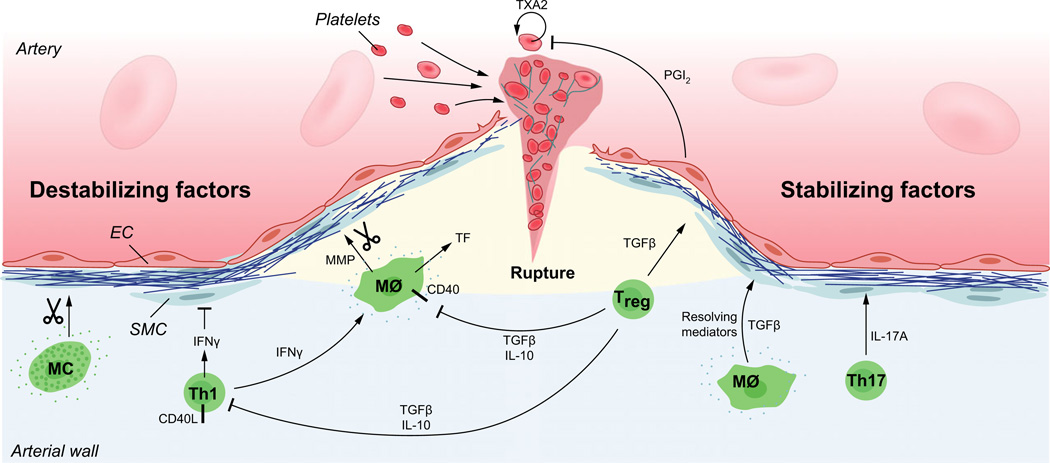
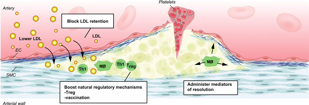
Atherosclerosis is a maladaptive, nonresolving chronic inflammatory disease that occurs at sites of blood flow disturbance. The disease usually remains silent until a breakdown of integrity at the arterial surface triggers the formation of a thrombus. By occluding the lumen, the thrombus or emboli detaching from it elicits ischaemic symptoms that may be life-threatening. Two types of surface damage can cause atherothrombosis: plaque rupture and endothelial erosion. Plaque rupture is thought to be caused by loss of mechanical stability, often due to reduced tensile strength of the collagen cap surrounding the plaque. Therefore, plaques with reduced collagen content are thought to be more vulnerable than those with a thick collagen cap. Endothelial erosion, on the other hand, may occur after injurious insults to the endothelium instigated by metabolic disturbance or immune insults. This review discusses the molecular mechanisms involved in plaque vulnerability and the development of atherothrombosis.

Keywords: atherosclerosis; atherothrombosis; en-dothelial erosion; inflammation; plaque rupture.


Figures

Fig. 1
Fig. 1
Mechanisms of plaque rupture. Activated macrophages and Th1 cells produce metalloproteinases and cytokines that hamper the tensile strength of the collagen cap. Several pro-inflammatory cytokines including interferon-Γ (IFNΓ) and tumour necrosis factor (TNF), as well as CD40/CD40L cell surface receptors of the TNF superfamily promote an inflammatory state that enhance cell death and prothrombotic activity in the plaque. When the cap no longer can withstand the mechanical force of the blood pressure, superficial fissures are formed in the plaque. Exposure of the plaque’s inner core with its thrombogenic material rapidly triggers platelet activation, humoral coagulation and the formation of a thrombus that may either occlude the artery at the site of plaque rupture or dissociate as an embolus and occlude the arterial lumen at a site downstream of the ruptured plaque. Counteracting all these pro-inflammatory and tissue-destructive signals, subsets of macrophages and T cells produce anti-inflammatory molecules that counteract vascular inflammation and reduce the risk for plaque rupture and atherothrombosis. Amongst them, transforming growth factor-β (TGF-β) and interleukin-10 (IL-10) inhibit inflammation and immune cell activation. In addition, TGF-β has fibrogenic properties that it shares with IL-17A produced by Th17 cells. The resolution of plaque inflammation depends not only on anti-inflammatory signals but also on resolving mediators such as eicosanoids of the resolvin type and Annexin I, both of which ligate the FPR/ALX receptor. EC, endothelial cell; SMC, smooth muscle cell; MΦ, macrophage; MMP, metalloproteinase; TXA2, thromboxane A2; PGI2, prostaglandin I2 (prostacyclin).
Fig. 2
Fig. 2
Mechanisms of plaque erosion. Endothelial cells of atherosclerotic plaques commonly express Toll-like receptor −2 (TLR2) that can ligate both Gram-positive toxins (G+ toxins) of bacterial pathogens and hyaluronan released from the extracellular matrix. TLR2 ligation can trigger endothelial dysfunction with endoplasmic reticulum stress and apoptosis. Such reactions are further enhanced by neutrophil attack on the endothelium. As a result, endothelial cells may detach, exposing the subendothelial matrix with its thrombogenic components. Activated neutrophils contribute to a prothrombotic state by releasing a set of proteases including neutrophil elastase and by forming neutrophil extracellular traps (NETs) that can damage endothelial cells, trap leucocytes and enhance thrombosis. PAD4, Peptide arginine deaminase-4, a component of NETs.
Fig. 3
Fig. 3
Therapy targets for prevention of atherothrombosis. Reduction of LDL (and other large lipoproteins) by lipid-lowering therapy and prevention of LDL retention in the artery wall, both act to reduce cholesterol accumulation, an initiator of atherosclerosis. Stimulation of immunoregulatory mechanisms reduces vascular inflammation; they include administration of anti-inflammatory cytokines, enhancing Treg cells and vaccination to elicit atheroprotective immunity. Mediators of resolution include resolvin-type eicosanoids, peptide mimetics of Annexin I and other substances.

References

    1. Tabas I, Garcia-Cardena G, Owens GK. Recent insights into the cellular biology of atherosclerosis. J Cell Biol. 2015;209:13–22. - PMC - PubMed
    1. Libby P, Ridker PM, Hansson GK. Progress and challenges in translating the biology of atherosclerosis. Nature. 2011;473:317–325. - PubMed
    1. Jonasson L, Holm J, Skalli O, Bondjers G, Hansson GK. Regional accumulations of t cells, macrophages, and smooth muscle cells in the human atherosclerotic plaque. Arteriosclerosis. 1986;6:131–138. - PubMed
    1. Hansson GK, Holm J, Jonasson L. Detection of activated t lymphocytes in the human atherosclerotic plaque. Am J Pathol. 1989;135:169–175. - PMC - PubMed
    1. Hansson GK, Hermansson A. The immune system in atherosclerosis. Nat Immunol. 2011;12:204–212. - PubMed

Publication types

MeSH terms

Substances