Review
. 2015 Aug 11:11:196.
doi: 10.1186/s12917-015-0515-5.

Canine atopic dermatitis: detailed guidelines for diagnosis and allergen identification

Affiliations
Review

Canine atopic dermatitis: detailed guidelines for diagnosis and allergen identification

Patrick Hensel et al. BMC Vet Res. .

Abstract

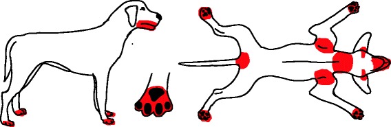
Background: Canine atopic dermatitis (AD) is a common, genetically predisposed, inflammatory and pruritic skin disease. The variation in clinical presentations, due to genetic factors, extent of the lesions, stage of the disease, secondary infections, as well as resemblance to other non-atopic related skin diseases, can complicate a diagnosis of canine AD. A sub-group of the International Committee for Allergic Diseases in Animals (ICADA) was tasked with the development of a set of practical guidelines that can be used to assist practitioners and researchers in the diagnosis of canine AD. Online citation databases and abstracts from international meetings were searched for publications related to the topic, and combined with expert opinion where necessary. The final set of guidelines was approved by the entire ICADA committee.

Results: A total of 81 publications relevant for this review were identified. The guidelines generated focus on three aspects of the diagnostic approach: 1. Ruling out of other skin conditions with clinical signs resembling, or overlapping with canine AD. 2. Detailed interpretation of the historical and clinical features of patients affected by canine AD. 3. Allergy testing by intradermal versus allergen-specific IgE serum testing.

Conclusions: The diagnosis of canine AD is based on meeting clinical criteria and ruling out other possible causes with similar clinical signs. Flea combing, skin scraping and cytology should be performed, where necessary, as part of a thorough work-up. Elimination diet trials are required for patients with perennial pruritus and/or concurrent gastrointestinal signs. Once a clinical diagnosis of canine AD is made, allergy testing can be performed to identify potential causative allergens for allergen-specific immunotherapy.


Figures

Fig. 1
Fig. 1
Distribution of skin lesions and pruritus associated with FAD. Acute lesions: Erythematous macules, papules, crusted papules, hot spots. Chronic lesions: Self-induced alopecia, lichenification, and hyperpigmentation
Fig. 2
Fig. 2
Distribution of skin lesions and pruritus associated with Lice/Cheyletiella. Lice: No visible lesions, or mild scaling and excoriation. Cheyletiella: Marked dorsal seborrhea
Fig. 3
Fig. 3
Distribution of skin lesions and pruritus associated with sarcoptic mange. Lesions include papular eruption, erythema, scaling, excoriations
Fig. 4
Fig. 4
Distribution of skin lesions and pruritus associated with trombiculiasis. Lesions usually manifest as eruption
Fig. 5
Fig. 5
Distribution of skin lesions and pruritus associated with otoacariasis. Lesions include erythema, dark-brown, coffee-ground like discharge
Fig. 6
Fig. 6
Distribution of skin lesions and pruritus associated with demodicosis. Lesions include focal, multi-focal or generalised alopecia, scaling, erythema, follicular casts, comedones, Furunculosis
Fig. 7
Fig. 7
Distribution of skin lesions and pruritus associated with Malassezia dermatitis. Lesions include erythema, yellowish or brownish greasy scale, hyperpigmentation
Fig. 8
Fig. 8
Common distribution of clinical lesions and pruritus associated with canine AD and food allergy
Fig. 9
Fig. 9
Silhouettes of atopic boxers, German shepherd dog, golden retrievers, shar peis, Dalmations, Labradors retriever, French bulldogs, West Highland white terriers and Jack Russell terriers (in this order). Each colour corresponds to the percentage of affected animals (Reproduced with permission from Veterinary Dermatology)
Fig. 10
Fig. 10
a, b, c Typical distribution of secondary skin lesions in a West Highland white terrier

References

    1. Halliwell R. Revised nomenclature for veterinary allergy. Vet Immunol Immunopathol. 2006;114(3-4):207–208. doi: 10.1016/j.vetimm.2006.08.013. - DOI - PubMed
    1. DeBoer DJ, Hillier A. The ACVD task force on canine atopic dermatitis (XV):fundamental concepts in clinical diagnosis. Vet Immunol Immunopathol. 2001;81(3-4):271–276. doi: 10.1016/S0165-2427(01)00312-9. - DOI - PubMed
    1. Wilhem S, Kovalik M, Favrot C. Breed-associated phenotypes in canine atopicdermatitis. Vet Dermatol. 2011;22(2):143–149. doi: 10.1111/j.1365-3164.2010.00925.x. - DOI - PubMed
    1. Nuttall T. The genomics revolution: will canine atopic dermatitis be predictable and preventable? Vet Dermatol. 2013;24(1):10–18. doi: 10.1111/j.1365-3164.2012.01094.x. - DOI - PubMed
    1. Favrot C, Steffan J, Seewald W, Picco F. A prospective study on the clinical featuresof chronic canine atopic dermatitis and its diagnosis. Vet Dermatol. 2010;21(1):23–31. doi: 10.1111/j.1365-3164.2009.00758.x. - DOI - PubMed

LinkOut - more resources