. 2015 Dec;149(7):1849-59.
doi: 10.1053/j.gastro.2015.07.062. Epub 2015 Aug 7.

Increased Expression of DUOX2 Is an Epithelial Response to Mucosal Dysbiosis Required for Immune Homeostasis in Mouse Intestine

Affiliations

Increased Expression of DUOX2 Is an Epithelial Response to Mucosal Dysbiosis Required for Immune Homeostasis in Mouse Intestine

Helmut Grasberger et al. Gastroenterology. 2015 Dec.

Erratum in

  • Correction.
    [No authors listed] [No authors listed] Gastroenterology. 2023 May;164(6):1033. doi: 10.1053/j.gastro.2023.02.020. Epub 2023 Mar 21. Gastroenterology. 2023. PMID: 36959022 No abstract available.

Abstract

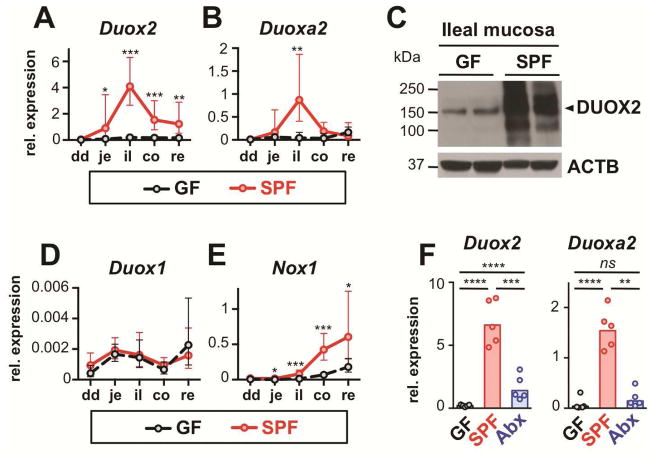
Background & aims: Dual oxidase 2 (DUOX2), a hydrogen-peroxide generator at the apical membrane of gastrointestinal epithelia, is up-regulated in patients with inflammatory bowel disease (IBD) before the onset of inflammation, but little is known about its effects. We investigated the role of DUOX2 in maintaining mucosal immune homeostasis in mice.

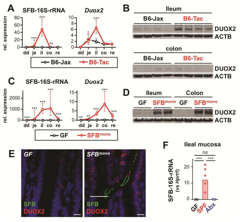
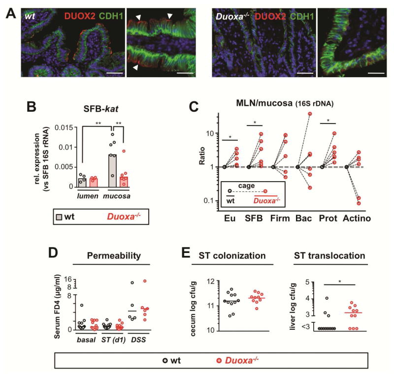
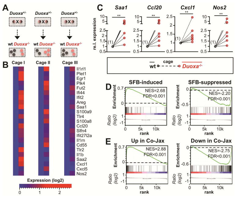
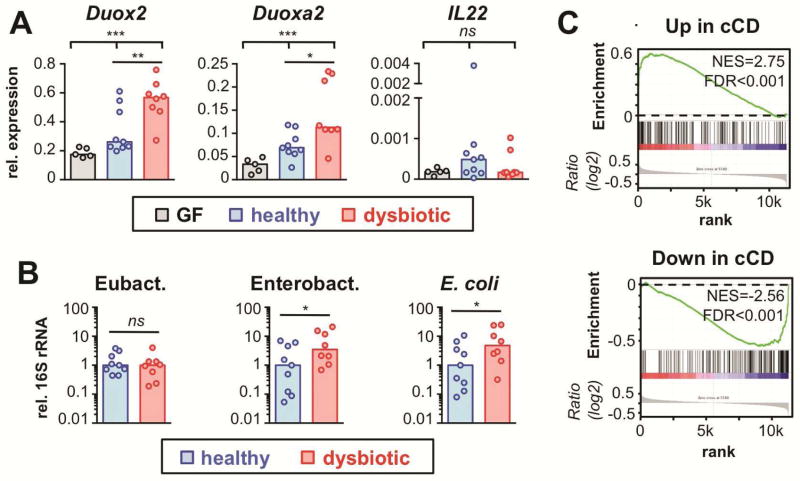
Methods: We analyzed the regulation of DUOX2 in intestinal tissues of germ-free vs conventional mice, mice given antibiotics or colonized with only segmented filamentous bacteria, mice associated with human microbiota, and mice with deficiencies in interleukin (IL) 23 and IL22 signaling. We performed 16S ribosomal RNA gene quantitative polymerase chain reaction of intestinal mucosa and mesenteric lymph nodes of Duoxa(-/-) mice that lack functional DUOX enzymes. Genes differentially expressed in Duoxa(-/-) mice compared with co-housed wild-type littermates were correlated with gene expression changes in early-stage IBD using gene set enrichment analysis.

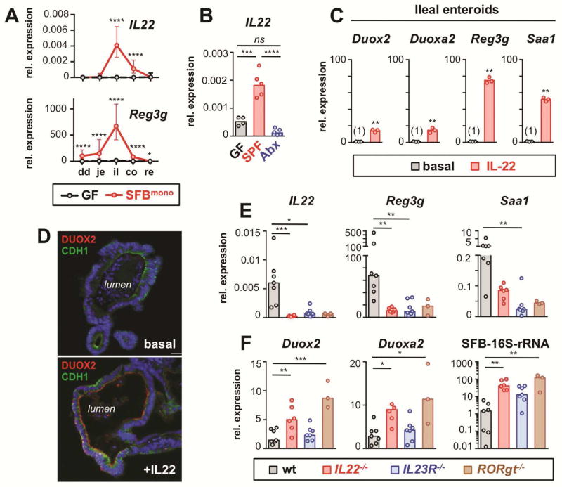
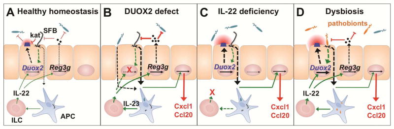
Results: Colonization of mice with segmented filamentous bacteria up-regulated intestinal expression of DUOX2. DUOX2 regulated redox signaling within mucosa-associated microbes and restricted bacterial access to lymphatic tissues of the mice, thereby reducing microbiota-induced immune responses. Induction of Duox2 transcription by microbial colonization did not require the mucosal cytokines IL17 or IL22, although IL22 increased expression of Duox2. Dysbiotic, but not healthy human microbiota, activated a DUOX2 response in recipient germ-free mice that corresponded to abnormal colonization of the mucosa with distinct populations of microbes. In Duoxa(-/-) mice, abnormalities in ileal mucosal gene expression at homeostasis recapitulated those in patients with mucosal dysbiosis.

Conclusions: DUOX2 regulates interactions between the intestinal microbiota and the mucosa to maintain immune homeostasis in mice. Mucosal dysbiosis leads to increased expression of DUOX2, which might be a marker of perturbed mucosal homeostasis in patients with early-stage IBD.

Keywords: Gastroenterology; Inflammatory Bowel Disease; Intestine; Microbial Dysbiosis.


Figures

Figure 1
Figure 1
Intestinal DUOX2 expression depends on microbial colonization. Relative Duox2 (A) and Duoxa2 (B) mRNA expression in GF (n=5) and SPF (n=7) mice. Data represent geometric means±95% CI. dd, duodenum; je, jejunum; il, ileum; co, colon; re, rectum. (C) Immunoblot of DUOX2 protein. ACTB, β-actin loading control. (D, E) Relative mRNA expression of Duox1 and Nox1. Data represent geometric means±95% CI. (F) Effect of acute enteral antibiotic treatment on ileal Duox2 and Duoxa2 expression. Mice were analyzed 24 hours following oral gavage with streptomycin (20 mg; Abx); SPF, sham treated control. Bars indicate geometric means. ****, P<.0001; ***, P<.001; **, P<.01; *, P<.05.
Figure 2
Figure 2
Mucosa-adherent SFB are a dominant inducer of ileal DUOX2 expression. (A) Mucosa-adherent SFB (16S-rRNA) and Duox2 mRNA level in SFBneg (B6-Jax; n=5) and SFBpos (B6-Tac; n=5) mice. Data represent geometric means±95% CI. dd, duodenum; je, jejunum; il, ileum; co, colon; re, rectum. (B) DUOX2 protein expression in ilea of B6-Jax and B6-Tac mice. Each lane represents an individual mouse. ACTB, β-actin loading control. (C, D) Mucosa-associated SFB and expression of Duox2 mRNA and protein in mice mono-associated for one week with SFB (SFBmono; n=5) or sham-treated GF controls (n=5). Data represent geometric means±95% CI. (E) Detection of DUOX2 protein by indirect immunofluorescence (red) and SFB by in situ hybridization (green) in the terminal ileum of GF and SFBmono mice. Scale bars, 10 μm. (F) Ileal mucosa-adherent SFB (16S-rRNA) in mice treated with oral streptomycin (Abx) or sham treated controls (SPF). ***, P<.001; **, P<.01; *, P<.05.
Figure 3
Figure 3
IL-22 can augment DUOX2 expression but is not essential for DUOX2 induction by microbial colonization. (A) Expression of IL22 and Reg3g along the intestinal tract of GF and SFBmono mice (n=5 per group). Data represent geometric means±95% CI. Note lower colonic expression of IL22 and Reg3g despite high level of mucosal SFB and DUOX2 (Fig 2C). (B) Acute microbial regulation of ileal IL22 expression. SPF (SFBpos) mice were analyzed 24 hours following oral gavage with streptomycin (Abx) or sham treatment (SPF). (C) Ileal enteroids were cultured with or without IL-22 (50 ng/ml for 18 h) and gene expression of selected genes determined by RT-qPCR. (D) Immunofluorescence of DUOX2 protein in IL-22-stimulated ileal enteroids. Cdh1, E-cadherin. (E) Ileal expression of IL22 and IL-22 target genes in wt and IL-22 deficient mice (IL22−/−, IL23R−/−, RORgt−/−). (F) Expression of DUOX2 subunit genes and mucosal SFB-colonization. Bars in panels E and F indicate median values. ****, P<.0001; ***, P<.001; **, P<.01; *, P<.05.
Figure 4
Figure 4
DUOX2 restricts transepithelial uptake of bacteria in the small intestine. (A) Detection of DUOX2 protein (red) at the epithelial brush border in the terminal ileum of wt but not Duoxa−/− littermates; the basolateral cell marker E-cadherin (CDH1) is shown in green; scale bars represent 50 μm (left panels) and 20 μm (right panels), respectively. (B) SFB-specific catalase (kat) expression in mucosa-adherent and luminal SFB. (C) Graph depicts the ratio of 16S rDNA in MLN vs ileal mucosa indicative of transepithelial bacterial flux. Dashed lines connect mean bacterial DNA level of Duoxa−/− mice and cohoused littermate controls. Eu, eubacteria; Firm, Firmicutes; Bac, Bacteroidetes; Prot, Gammaproteobacteria; Actino, Actinobacteria. (D) In vivo intestinal permeability for 4 kDa dextran in Duoxa−/− and wt mice at baseline, following acute enteral Salmonella infection (ST), and after treatment with dextran sulfate sodium (DSS) to induce unspecific epithelial injury. (E) Acute systemic dissemination of enteral Salmonella Typhimurium in Duoxa−/− and wt animals 24 h post enteral infection.
Figure 5
Figure 5
Loss of DUOX activity disturbs mucosal homeostasis in the terminal ileum. (A) Experimental setup for gene expression profiling. (B) Expression heat maps depicting selected genes upregulated in the ileum of Duoxa−/− animals. (C) Ileal gene expression analyzed by RT-qPCR. Mean expression in Duoxa−/− animals is plotted relative to the mean in cohoused wt littermates (set to 1) in cage-wise comparisons (n=7). **, P<.01. (D, E) Genes controlled by SFB-monocolonization or by cohousing of B6-Jax (SFBneg) with B6-Tac (SFBpos) mice were tested for correlation with genes affected by loss of DUOX activity using GSEA (see Supplementary Methods). Within each plot, genes are sorted for their relative ileal expression in Duoxa−/− mice (left side: up in Duoxa−/−; right side: down in Duoxa−/−). Genes upregulated (downregulated) by introduction of SFB are significantly correlated with those upregulated (downregulated) in mice with loss of DUOX activity. Core enriched genes are listed in Supplementary Tables S16-S19. FDR, false discovery rate q-value; NES, normalized enrichment score.
Figure 6
Figure 6
DUOX2 is induced by constituents of the microbiota from IBD patients and acts as a compensatory mucosal defense pathway. (A) Gene expression in colonic mucosa of mice colonized for two weeks with fecal material from healthy donors or patients with ulcerative colitis-associated dysbiosis. Bars indicate median values. Kruskal-Wallis test; ***, P<.001; **, P<.01; *, P<.05. (B) 16S rRNA level in mucosal samples corresponding to (A). *, P<.05. (C) Genes dysregulated in the non-affected ileum of patients with colon-only CD (cCD vs healthy controls) were tested for correlation with genes affected by loss of DUOX2 activity using GSEA. Genes upregulated (downregulated) in non-inflamed tissue from CD patients are significantly enriched among those upregulated (downregulated) in mice with loss of DUOX activity. Leading edge gene subsets are depicted in Tables S20 and S21. FDR, false discovery rate q-value; NES, normalized enrichment score.
Figure 7
Figure 7
Model for the integration of the DUOX2 system into the intestinal epithelial defense response. (A) Epithelial contacting microbes (e.g., SFB) induce DUOX2 by an IL-22-independent pathway at homeostasis. DUOX2 activity triggers an anti-oxidative response (kat) in mucosa-adherent SFB but does not prevent their colonization. (B) Lack of DUOX2 activity leads to increased uptake of bacterial material (e.g., SFB, Proteobacteria). Expression of compensatory host defense systems resulting in a proinflammatory milieu. (C) Lack of IL-22 dependent mucosal defense leads to mucosal dysbiosis and compensatory induction of DUOX2. (D) In IBD-associated mucosal dysbiosis, increased epithelial contact with and uptake of indigenous pathobionts triggers a compensatory DUOX2 response before the onset of clinical inflammation. Sensing of bacterial factors by antigen-presenting cells (APC) activates the IL-23/IL-22 cascade for secretion of antimicrobial effectors and further enhancement of DUOX2 expression.

References

    1. Goto Y, Ivanov II. Intestinal epithelial cells as mediators of the commensal-host immune crosstalk. Immunol Cell Biol. 2013;91:204–14. - PMC - PubMed
    1. Geiszt M, Witta J, Baffi J, et al. Dual oxidases represent novel hydrogen peroxide sources supporting mucosal surface host defense. FASEB J. 2003;17:1502–4. - PubMed
    1. El Hassani RA, Benfares N, Caillou B, et al. Dual oxidase2 is expressed all along the digestive tract. American journal of physiology Gastrointestinal and liver physiology. 2005;288:G933–42. - PubMed
    1. Ha EM, Oh CT, Bae YS, et al. A direct role for dual oxidase in Drosophila gut immunity. Science. 2005;310:847–50. - PubMed
    1. Chavez V, Mohri-Shiomi A, Garsin DA. Ce-Duox1/BLI-3 generates reactive oxygen species as a protective innate immune mechanism in Caenorhabditis elegans. Infection and immunity. 2009;77:4983–9. - PMC - PubMed

Publication types

MeSH terms