Review
. 2015 Aug 10;8(3):a019232.
doi: 10.1101/cshperspect.a019232.

Mechanical Forces and Growth in Animal Tissues

Affiliations
Review

Mechanical Forces and Growth in Animal Tissues

Loïc LeGoff et al. Cold Spring Harb Perspect Biol. .

Abstract

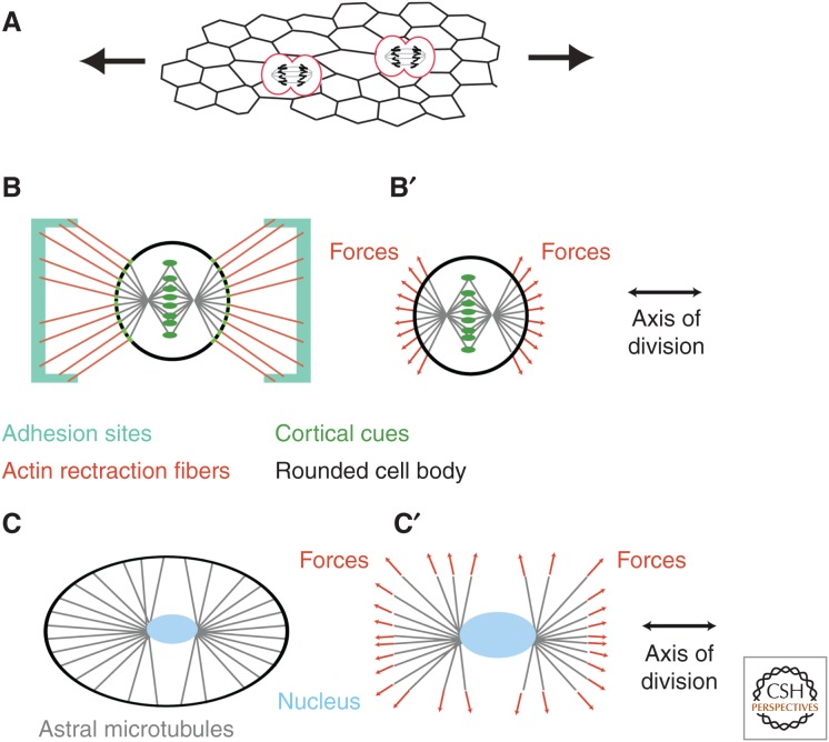
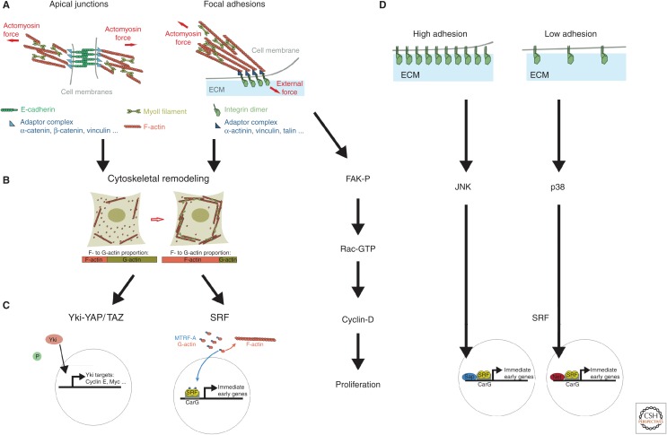
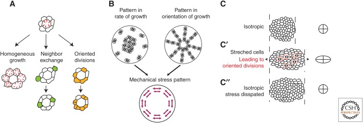
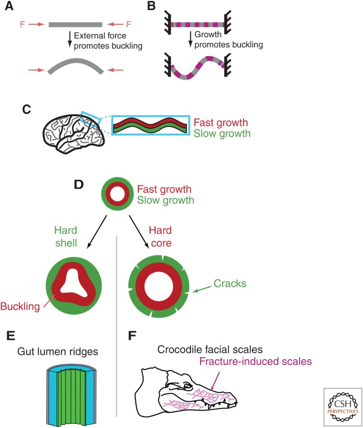
Mechanical forces shape biological tissues. They are the effectors of the developmental programs that orchestrate morphogenesis. A lot of effort has been devoted to understanding morphogenetic processes in mechanical terms. In this review, we focus on the interplay between tissue mechanics and growth. We first describe how tissue mechanics affects growth, by influencing the orientation of cell divisions and the signaling pathways that control the rate of volume increase and proliferation. We then address how the mechanical state of a tissue is affected by the patterns of growth. The forward and reverse interactions between growth and mechanics must be investigated in an integrative way if we want to understand how tissues grow and shape themselves. To illustrate this point, we describe examples in which growth homeostasis is achieved by feedback mechanisms that use mechanical forces.


Figures

Figure 1.
Figure 1.
Orienting cell divisions. (A) The existence of external forces in a tissue induces internal stresses that deform cells. This deformation may then orient cells divisions. (B) In cells adhering to a substrate, experiments on isolated cells cultured on patterned substrates have shown that the rounded cell body may keep the memory of its adherent geometry through actin-rich retraction fibers, which pull on the cell cortex. (B′) Somehow, the pulling forces are transmitted to the astral microtubules, which results in orienting the mitotic spindle. (C) Observations of unperturbed embryos, as well as recent experiments in confined geometries, have highlighted how cells sense their shape to orient the mitotic spindle. (C′) The shape of cells can be sensed by astral microtubules, which extend to the cell cortex and generate a global torque and force to orient the centrosomes and nucleus, or spindle, depending on whether the alignment occurs before or after nuclear envelope breakdown.
Figure 2.
Figure 2.
Mechanical signaling and tissue growth. (A) Force generation and force transmission occur at apical adherens junctions (left) and basal focal adhesion sites (right). At apical junctions, the contractile actomyosin cortices of two neighboring cells are mechanically coupled via the E-cadherin adhesion complex. The adaptor complex, which links E-cadherin to the cortex, includes proteins, such as vinculin, which is mechanosensitive. At basal sites of adhesion, force balance between the contractile actomyosin and the ECM is sensed at sites of focal adhesion, which involves integrin dimers and associated proteins coupling to actin filaments. (B) Force sensation is then converted into a structural reorganization of the actin cytoskeleton, increased external force promoting polymerized actin. (C) The polymerized state of the actin cytoskeleton is then sensed by the transcription activators YAP/TAZ-Yki, a downstream component from the Hippo pathway– or myocardin-related transcription factor (MRTF)-A, a component of the serum response factor (SRF) pathway, which both shuttle to the nucleus to promote growth. (D) Mechanical signaling can also affect growth in the absence of cytoskeletal reorganization. Force sensation at focal adhesions leads to the phosphorylation of the focal adhesion kinase (FAK), which induces expression of cyclin D. The SRF pathway has also been proposed to sense the level of adhesion with the ECM in an actin-independent way, via Jun amino-terminal kinase (JNK) and p38. Also involved in this regulation and shown on the figure are the SRF cofactors (Sap and Net) at the SRF-binding consensus element (CArG).
Figure 3.
Figure 3.
How cell growth impacts on tissue mechanics. (A) If a cell grows (red arrows) more than its neighbors, a mechanical stress is generated that results in increased pressure in the growing cell and the stretching of its neighbors (top). The stress disappears when the neighbors also grow until growth is spatially homogeneous (left). Other mechanisms that can dissipate the mechanical stress are cell exchange (see the green cells in the middle), and oriented divisions along the axis of stress (see the orange cells on the right). (B) Patterns of growth, such as growth heterogeneities or polarized growth, induce mechanical stresses in a tissue. Regional differences in tissue growth (left) may lead to mechanical stress in the tissue. Here, growth was symbolized by cell division, but it is indeed the volume increase that matters, not cell divisions. If more growth proceeds in the center of the tissue, and provided tissue is sufficiently elastic to prevent stress dissipation, this will result in a stretch of the periphery of the tissue. Oriented cell division (right) can lead to similar patterns of mechanical stress, provided they are accompanied by volume increase. (C) In a tissue with no preferred direction, cells are isotropic, and the average fit to an ellipse yields a circle (right). (C′) An external uniaxial mechanical stress (which may originate from neighboring regions) deforms cells along its axis (right: average fit to an ellipse is elongated). This stretch promotes orientation of cell divisions (red dividing cells on the schematic). (C″) As a result of the polarized growth, cells will at least partially lose their stretch (averaged fit to an ellipse is a circle). Thus, oriented cell divisions contribute to the dissipation of the mechanical stress that was imposed on the tissue.
Figure 4.
Figure 4.
Making 3D shapes. (A) If an elastic rod or sheet is subjected to compressive forces, the rod will buckle above a certain threshold. (B) Buckling may also proceed in the absence of an external force when the structure grows, provided it is constrained at its ends. (C) Folds of the mammalian cortex have been proposed to stem from a buckling instability in which the fast growth of the external layer of the cortex is constrained by the slower growth of another layer below it. (DF) Apposition of tissues with different growth rates may give rise to very different 3D structures depending on the boundary conditions. To illustrate this, we consider a fast-growing layer surrounded by a slow-growing layer. If the surrounding layer is rigid and makes a hard shell (left), buckling may proceed in the fast-growing inner layer. Such a mechanism has been proposed to initiate gut lumen ridges (E). If it is the fast-growing layer that is the strongest and makes a hard core (right), then the external layer will be deformed and may even crack. This has been proposed to drive the formation of facial scales of crocodiles (F).

References

    1. Aegerter-Wilmsen T, Aegerter CM, Hafen E, Basler K. 2007. Model for the regulation of size in the wing imaginal disc of Drosophila. Mech Dev 124: 318–326. - PubMed
    1. Aigouy B, Farhadifar R, Staple DB, Sagner A, Röper J-C, Jülicher F, Eaton S. 2010. Cell flow reorients the axis of planar polarity in the wing epithelium of Drosophila. Cell 142: 773–786. - PubMed
    1. Ambegaonkar AA, Pan G, Mani M, Feng Y, Irvine KD. 2012. Propagation of Dachsous-Fat planar cell polarity. Curr Biol 22: 1302–1308. - PMC - PubMed
    1. Aragona M, Panciera T, Manfrin A, Giulitti S, Michielin F, Elvassore N, Dupont S, Piccolo S. 2013. A mechanical checkpoint controls multicellular growth through YAP/TAZ regulation by actin-processing factors. Cell 154: 1047–1059. - PubMed
    1. Baena-Lopez LA, Baonza A, Garcia-Bellido A. 2005. The orientation of cell divisions determines the shape of Drosophila organs. Curr Biol 15: 1640–1644. - PubMed

Publication types

LinkOut - more resources