. 2015 Aug 25;112(34):10774-9.
doi: 10.1073/pnas.1504276112. Epub 2015 Aug 10.

The amino acid sensor GCN2 inhibits inflammatory responses to apoptotic cells promoting tolerance and suppressing systemic autoimmunity

Affiliations

The amino acid sensor GCN2 inhibits inflammatory responses to apoptotic cells promoting tolerance and suppressing systemic autoimmunity

Buvana Ravishankar et al. Proc Natl Acad Sci U S A. .

Abstract

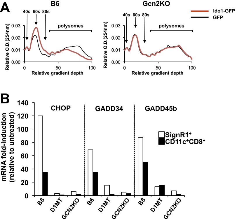
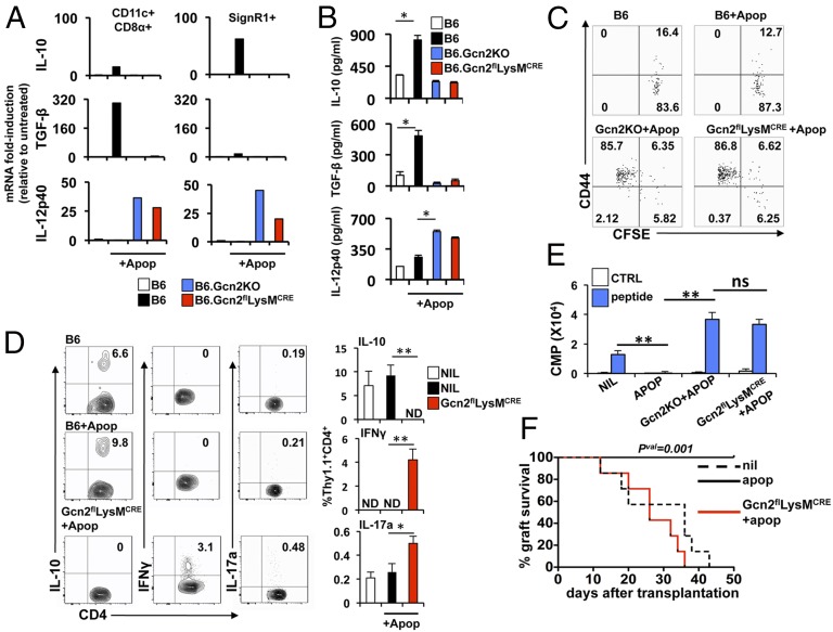
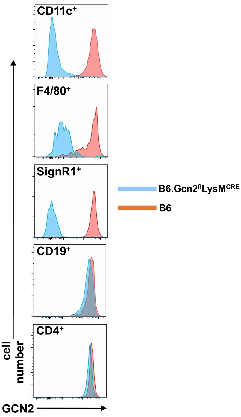
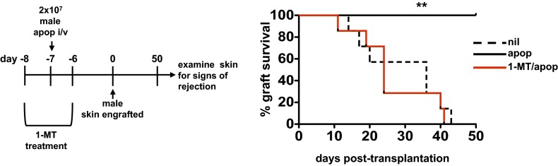
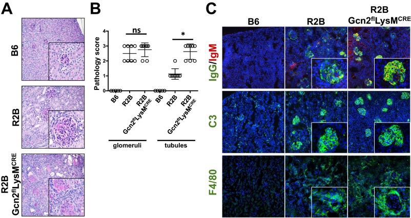
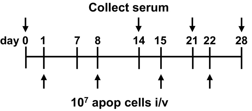
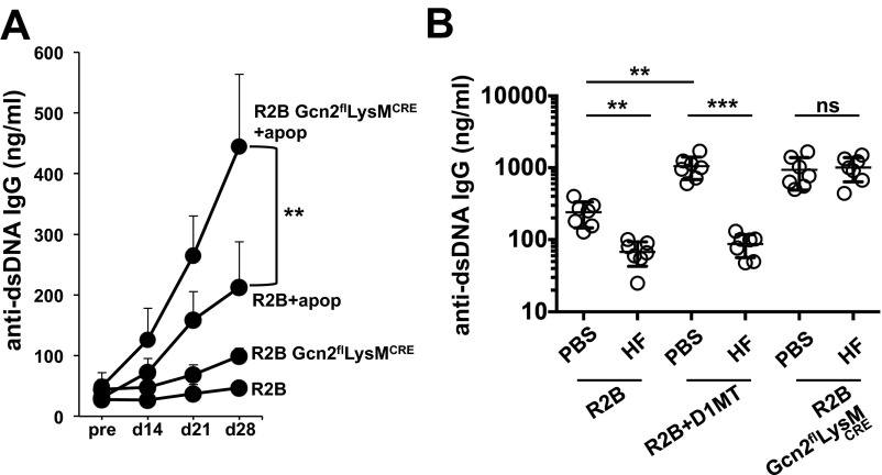
Efficient apoptotic cell clearance and induction of immunologic tolerance is a critical mechanism preventing autoimmunity and associated pathology. Our laboratory has reported that apoptotic cells induce tolerance by a mechanism dependent on the tryptophan catabolizing enzyme indoleamine 2,3 dioxygenase 1 (IDO1) in splenic macrophages (MΦ). The metabolic-stress sensing protein kinase GCN2 is a primary downstream effector of IDO1; thus, we tested its role in apoptotic cell-driven immune suppression. In vitro, expression of IDO1 in MΦs significantly enhanced apoptotic cell-driven IL-10 and suppressed IL-12 production in a GCN2-dependent mechanism. Suppression of IL-12 protein production was due to attenuation of IL-12 mRNA association with polyribosomes inhibiting translation while IL-10 mRNA association with polyribosomes was not affected. In vivo, apoptotic cell challenge drove a rapid, GCN2-dependent stress response in splenic MΦs with increased IL-10 and TGF-β production, whereas myeloid-specific deletion of GCN2 abrogated regulatory cytokine production with provocation of inflammatory T-cell responses to apoptotic cell antigens and failure of long-tolerance induction. Consistent with a role in prevention of apoptotic cell driven autoreactivity, myeloid deletion of GCN2 in lupus-prone mice resulted in increased immune cell activation, humoral autoimmunity, renal pathology, and mortality. In contrast, activation of GCN2 with an agonist significantly reduced anti-DNA autoantibodies and protected mice from disease. Thus, this study implicates a key role for GCN2 signals in regulating the tolerogenic response to apoptotic cells and limiting autoimmunity.

Keywords: apoptosis; autoimmunity; immunometabolism; stress; tolerance.


Conflict of interest statement

The authors declare no conflict of interest.

Figures

Fig. 1.
Fig. 1.
GCN2 activation promotes apoptotic cell-driven IL-10 production in MΦs. (A and B) BM MΦs were transduced with IDO1-GFP+ or control lentivirus as described in Materials and Methods. Then, 24 h later, kynurenine and Trp in the culture supernatant mRNA for the indicated genes were measured by sqPCR. (C) At 24 h after lentiviral transduction as described in A, BM MΦs were incubated with apoptotic thymocytes at a 10:1 apoptotic cell:MΦ ratio. Then, 18 h later, cell supernatants were collected, and cytokines were measured by ELISA. (D) Western blot analysis for the proteins indicated was done on whole-cell lysates from BM MΦ cultures as described in C. (E) mRNA from BM MΦ/apoptotic cell cultures as in C was collected at the indicated time points, and cytokine message expression was assessed by sqPCR. In some groups, 20 μM D1MT was added. (F) Sucrose gradient fractionation of cellular lysates was followed by sqPCR analysis for the indicated mRNA species from 8-h BM MΦ/apoptotic cultures treated as in C. In A, B, C, and E, bars and line points represent mean ± SD values for triplicate samples. In D, Western blot images are representative of three separate experiments. In F, pooled material from triplicate samples was analyzed. *P < 0.05; **P < 0.01, Student’s t test. Experiments were repeated three times, with similar results.
Fig. S1.
Fig. S1.
Semiquantative assessment of Western blot band intensity. (A and B) Relative band intensity for phospho-eIF2α (A) normalized to total eIF2α and total ATF4 (B) normalized to β-actin for blots shown in Fig. 1D. (C and D) Relative band intensity for phospho-eIF2α (C) normalized to total eIF2α and total IDO1 (D) normalized to β-actin for blots shown in Fig. 3B. Bars represent mean ± SD values for triplicate samples. Experiments were repeated three times, with similar results.
Fig. S2.
Fig. S2.
IDO1 expression decreases RNA association with polysomes in vitro, and apoptotic cell challenge induces IDO1- and GCN2-dependent stress gene expression in MZ MΦs and CD8α+ DCs in vivo. (A) Polysome analysis of IDO1+ MΦs incubated with apoptotic cells as described in Fig. 1. Cell extracts were subjected to sucrose density centrifugation, and RNA association with polysomes was analyzed by measuring the absorbance at 254 nM. Ribosomal peaks at 40 s, 60 s, and 80 s are identified. (B) At 4 h after apoptotic cell challenge (107 in vivo), SignR1+ MZ MΦs and CD11c+CD8α+ DCs were sorted by FACS from the spleen, and message expression for the indicated mRNA species was determined by sqPCR. Bars represent the relative induction vs. controls value for pooled samples from four mice. In one experimental condition, mice were given the IDO1 inhibitor D1MT ad libitum in drinking water as described in SI Materials and Methods. The experiment was repeated four times, with similar results.
Fig. 2.
Fig. 2.
Myeloid GCN2 signals are required to suppress adaptive immunity to apoptotic cells in vivo. (A) At 4 h after apoptotic cell challenge (107 in vivo), SignR1+ MZ MΦs and CD11c+CD8α+ DCs were sorted by FACS, and cytokine message expression for the indicated species was determined by sqPCR. (B) Mice of the indicated phenotype were challenged with 107 apoptotic cells; 18 h later, spleens were collected, and cytokine protein concentrations were determined on whole spleen lysate by ELISA. (C) Mice received 2.5 × 10⁶ MACS-purified CFSE-labeled Thy1.1+CD4+ OTII T cells in vivo, followed by challenge with 107 OVA+ apoptotic cells. At 3 d later, spleens were collected, and CFSE and CD44 intensity in the CD4+Thy1.1+ T cells was determined by flow cytometry. Plots are gated on Thy1.1+CD4+ cells. (D) Splenocytes from mice treated as described in C were restimulated in vitro for 5 h with PMA/ionomycin as described in SI Materials and Methods, after which intracellular FACS analysis for the indicated cytokines in Thy1.1+OTII+ T cells was done. (E) FACS-purified Thy1.1+OTII+ T cells were stimulated with peptide-pulsed CD11c+ DCs, and proliferation was determined by thymidine incorporation. cpm, count per minute. (F) Male B6 thymocytes (107) were adoptively transferred in vivo into mice of the indicated genotype, followed by male skin engraftment 7 d later (as described in SI Materials and Methods), and then monitored for graft rejection for 50 d. n = 10 mice/group. In A, bars represent the value for pooled samples from four mice. In B and D, bars represent mean ± SD values for five mice. In C and D, plots are representative of five mice per group. *P < 0.05, **P < 0.01, Student’s t test. Experiments were repeated four times, with similar results.
Fig. S3.
Fig. S3.
FACS staining for intracellular GCN2 in splenocytes from B6.Gcn2flLysMCRE mice. Shown are representative histograms of splenic cell populations identified with the indicated markers stained for the intracellular presence of GCN2, as described in SI Materials and Methods. The experiment was repeated twice, with the same results. n = 4 mice per group.
Fig. S4.
Fig. S4.
IDO1 activity at the time of apoptotic cell exposure is required for generation of stable tolerance and allograft acceptance. Female B6 mice were given the IDO1 inhibitor D1MT ad libitum in drinking water for 2 d before administration of 107 male B6 thymocytes in vivo. Mice were then immediately taken off the D1MT containing water, followed by male skin engraftment 7 d later (as described in SI Materials and Methods), and monitored for graft rejection for 50 d. The experiment was repeated twice, with similar results. n = 10 mice/group.
Fig. 3.
Fig. 3.
MZ MΦ and B-cell activation are increased in lupus-prone mice lacking myeloid GCN2 function. (A) Splenocyte numbers from 6-mo-old mice of the indicated genotype. (B) Representative Western blot of whole spleen lysate from two mice per group described in A. m, mouse. (C–E) Flow cytometry analysis of groups described in A. In D, B-cell plots shown are gated on a B220+ population. Graphs in E are gated on the markers indicated above. Bars represent mean ± SD values for eight mice. *P < 0.05, **P < 0.01, Student’s t test. ns, not significant. Experiments were repeated four times, with similar results.
Fig. 4.
Fig. 4.
GCN2 limits inflammatory cytokine production and systemic autoimmune disease in lupus-prone mice. (A) SignR1+ MZ MΦs were sorted from 6-mo-old mice of the indicated genotype by FACS and cytokine message for the indicated species was determined by sqPCR. (B) Whole spleen lysates from 6-mo-old mice of the indicated genotype were collected, and cytokine protein concentrations were determined by ELISA. (C) Total serum IgG from 6-mo-old mice was measured by ELISA. (D) Serum was collected monthly from mice at the indicated ages, and IgG reactivity against dsDNA was measured by ELISA. (E) Urine was collected from 6- to 7- mo-old mice, and protein concentrations were measured as described in Materials and Methods. (F) Survival for mice of the indicated genotype over a 12-mo period was monitored. In A, bars represent the value for pooled samples from four mice. In B and C, bars represent mean ± SD values for eight mice. In D and E, graphs show mean ± SD values for eight mice. In F, n = 10 mice per group, and significance was determined as described in Materials and Methods. *P < 0.05, **P <0.01, ***P < 0.0001, Student’s t test. ns, not significant. Experiments were repeated three times, with similar results, with the exception of the experiment shown in F, which was repeated twice.
Fig. S5.
Fig. S5.
Renal pathology is increased in lupus-prone mice with deficiencies in myeloid GCN2 function. Kidneys were collected from 6-mo-old female mice of the indicated genotype. (A) Representative PAS-stained sections from the mice. (B) Tubular and glomerular pathology scores from mice in A determined as described in SI Materials and Methods. (C) Kidney cryosections from mice were stained for for IgG, IgM, complement C3, and F4/80. n = 6 mice/group. (Scale bar: 100 μm.)
Fig. S6.
Fig. S6.
Schematic for the experimental approach in Fig. S7A.
Fig. S7.
Fig. S7.
Autoimmunity is accelerated in myeloid GCN2KO mice exposed to apoptotic cells. (A) 3-mo-old mice of the indicated genotype were injected with 107 B6 apoptotic cells once weekly (four times total). Serum was collected at the indicated time points, and concentrations of total IgG reactive against dsDNA as a marker of systemic autoimmunity were determined by ELISA. (B) R2B and R2B GCN2flLysMcre mice were given HF injections i.p. as described in SI Materials and Methods for a period of 2 mo, after which serum was collected and IgG reactivity to dsDNA was measured by ELISA. In one experimental condition, mice were given the IDO1 inhibitor D1MT ad libitum in drinking water and examined for the impact on autoimmune disease with and without HF treatment. For both experiments, n = 7 mice/group. *P < 0.05, **P < 0.01, Student’s t test. ns, not significant. Experiments were repeated three times, with similar results.

References

    1. Casciola-Rosen LA, Anhalt G, Rosen A. Autoantigens targeted in systemic lupus erythematosus are clustered in two populations of surface structures on apoptotic keratinocytes. J Exp Med. 1994;179(4):1317–1330. - PMC - PubMed
    1. Rosen A, Casciola-Rosen L. Autoantigens as substrates for apoptotic proteases: Implications for the pathogenesis of systemic autoimmune disease. Cell Death Differ. 1999;6(1):6–12. - PubMed
    1. Colonna L, Lood C, Elkon KB. Beyond apoptosis in lupus. Curr Opin Rheumatol. 2014;26(5):459–466. - PMC - PubMed
    1. McGaha TL, Chen Y, Ravishankar B, van Rooijen N, Karlsson MC. Marginal zone macrophages suppress innate and adaptive immunity to apoptotic cells in the spleen. Blood. 2011;117(20):5403–5412. - PubMed
    1. Ravishankar B, McGaha TL. O death where is thy sting? Immunologic tolerance to apoptotic self. Cell Mol Life Sci. 2013;70(19):3571–3589. - PMC - PubMed

Publication types

MeSH terms