Comparative Study
. 2015 Aug 25;112(34):10798-803.
doi: 10.1073/pnas.1503129112. Epub 2015 Aug 10.

The opportunistic marine pathogen Vibrio parahaemolyticus becomes virulent by acquiring a plasmid that expresses a deadly toxin

Affiliations
Comparative Study

The opportunistic marine pathogen Vibrio parahaemolyticus becomes virulent by acquiring a plasmid that expresses a deadly toxin

Chung-Te Lee et al. Proc Natl Acad Sci U S A. .

Erratum in

Abstract

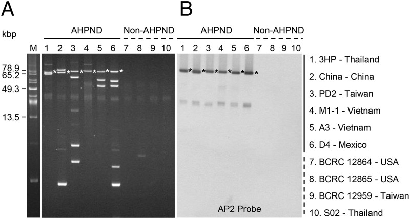
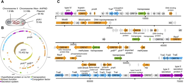
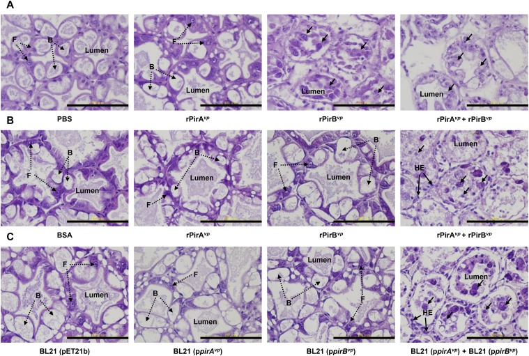
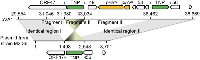
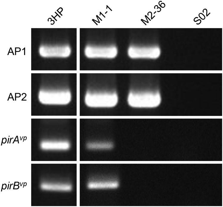
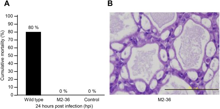
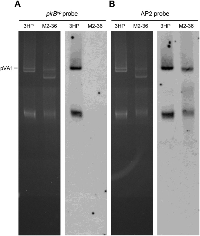
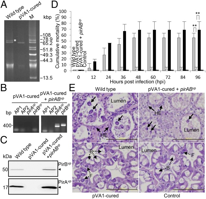
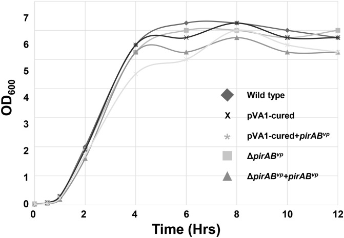
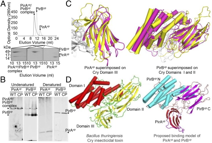
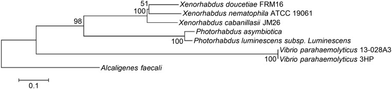
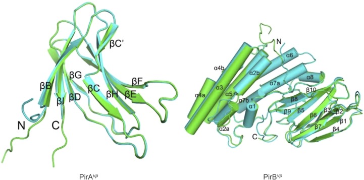
Acute hepatopancreatic necrosis disease (AHPND) is a severe, newly emergent penaeid shrimp disease caused by Vibrio parahaemolyticus that has already led to tremendous losses in the cultured shrimp industry. Until now, its disease-causing mechanism has remained unclear. Here we show that an AHPND-causing strain of V. parahaemolyticus contains a 70-kbp plasmid (pVA1) with a postsegregational killing system, and that the ability to cause disease is abolished by the natural absence or experimental deletion of the plasmid-encoded homologs of the Photorhabdus insect-related (Pir) toxins PirA and PirB. We determined the crystal structure of the V. parahaemolyticus PirA and PirB (PirA(vp) and PirB(vp)) proteins and found that the overall structural topology of PirA(vp)/PirB(vp) is very similar to that of the Bacillus Cry insecticidal toxin-like proteins, despite the low sequence identity (<10%). This structural similarity suggests that the putative PirAB(vp) heterodimer might emulate the functional domains of the Cry protein, and in particular its pore-forming activity. The gene organization of pVA1 further suggested that pirAB(vp) may be lost or acquired by horizontal gene transfer via transposition or homologous recombination.

Keywords: AHPND; Pir toxin; Vibrio parahaemolyticus; shrimp; virulence plasmid.


Conflict of interest statement

The authors declare no conflict of interest.

Figures

Fig. 1.
Fig. 1.
Analysis of plasmids purified from AHPND and non-AHPND strains. (A) Uncut plasmids from various AHPND-causing strains (lanes 1–6) and non-AHPND strains (lanes 7–10) after separation in 0.8% agarose gel. The size marker lane (marked “M”) shows uncut plasmids from Pantoea stewartii SW2 (29). (B) Southern blot hybridization signals of the same uncut plasmids after they were transferred to a nylon membrane and probed with alkaline phosphatase-labeled DNA fragments derived from the AP2 amplicon. Asterisks indicate corresponding plasmid bands.
Fig. S1.
Fig. S1.
Full sequence of the AHPND-associated plasmid pVA1. (A) Genomic material in the V. parahaemolyticus strain 3HP. Circular (B) and linear (C) diagrams of the plasmid pVA1 generated by DNAPlotter (42). ORFs were predicted by RAST (15) and annotated manually by BlastP using the National Center for Biotechnology Information Web site. In B, the innermost circle shows a GC plot, with dark yellow representing below-average GC content and brown indicating above-average GC content.
Fig. 2.
Fig. 2.
PirABvp toxin leads to shrimp HP cell death, the characteristic sloughing of the dead epithelial cells into the HP tubules, and mortality within 24 h. (A) Western blot analysis with anti-PirAvp and anti-PirBvp antibodies detected PirAvp and PirBvp proteins in the medium of a V. parahaemolyticus culture during the log phase of growth (1–4 h). (B) PCR confirmation of the AP1, AP2, pirAvp, and pirBvp sequences in the ΔpirABvp and ΔpirABvp+pirABvp strains. (C) Western blot analysis with anti-PirAvp and anti-PirBvp antibodies shows the presence or absence of PirAvp and PirBvp proteins in the ΔpirABvp and ΔpirABvp+pirABvp strains. (D) Virulence assay shows cumulative mortalities of shrimp immersed in the WT, ΔpirABvp, and the complemented strains. Shrimp immersed in TSB were used as a negative control. Significant differences were determined by unpaired Student t test (**P< 0.005). (E) Sections of the HP from shrimp in the virulence assay were stained with H&E and subjected to histological examination. The typical AHPND signs of sloughed epithelial cells (arrows) and hemocytic encapsulation (HE) were induced by the WT and the complemented strains, whereas shrimp immersed in the ΔpirABvp strain or the TSB control exhibited normal B- and F-cells (broken arrows) without any sloughing of the tubule cells. (Scale bar: 100 μm.) (F) To confirm the toxin content used in the per os challenge, Western blot with anti-PirA and anti-PirB probes was used to detect the purified recombinant Pir toxins rPirAvp, rPirBvp, rPirAvp+rPirBvp, and the E. coli recombinants BL21 (ppirAvp), BL21 (ppirBvp), and BL21 (ppirAvp) + BL21 (ppirBvp). WT AHPND strain 3HP was used as the positive control, and BSA and the empty vector BL21 (pET21b) were used as the negative controls. (G and H) Results of per os challenge show cumulative mortalities of shrimp fed with pellets soaked for 10 min in solutions containing (G) purified recombinant Pir toxin rPirAvp, rPirBvp, and rPirAvp+rPirBvp and (H) the E. coli recombinants BL21 (ppirAvp), BL21 (ppirBvp), and BL21 (ppirAvp) + BL21 (ppirBvp). Shrimp fed with pellets soaked in BSA or in the E. coli recombinant with the expression vector only, BL21 (pET21b), was used as the negative controls. Significant differences were determined by unpaired Student t test (**P< 0.005).
Fig. S2.
Fig. S2.
H&E-stained sections of the HP from shrimp challenged by reverse gavage (A) and per os (B and C). For reverse gavage, shrimp were injected with PBS solution or 100 μL 2 μM rPirAvp, rPirBvp, or premixed rPirAvp/rPirBvp. Normal B- and F-cells without any sloughing of the tubule cells were seen in the shrimp injected with PBS solution. Injection of rPirAvp+rPirBvp or PirBvp produced epithelial sloughing (arrows). For the per os feeding challenge, after 1 d of starvation, shrimp were fed with pellets soaked in the indicated protein(s) or E. coli recombinant(s). Epithelial sloughing (arrows) was observed only when challenged with pellets containing both rPirAvp and rPirBvp or with E. coli BL21 recombinants that expressed both rPirA and rPirB. (Scale bar: 100 μm.)
Fig. 3.
Fig. 3.
Comparison of pVA1 and the non–AHPND-causing plasmid from strain M2-36 shows the location of the missing DNA fragments. Putative pVA1 ORFs are indicated by the boxes marked with arrows. In the sequence amplified by the AP1-F16/AP2-R1 primer set (Table S2), fragments I and III are absent from the plasmid in the M2-36 strain, whereas fragment II is in reverse orientation. The blue arrowheads represent inverted repeats.
Fig. S3.
Fig. S3.
PCR detection of the AP1, AP2, pirAvp, and pirBvp sequences in AHPND and non-AHPND strains by PCR. PCR was performed on the indicated strains after they had been cultured from stock for 2 h at 37 °C with agitation. The PCR products were then separated by electrophoresis in a 2.0% (wt/vol) agarose gel, and the appearance of a band of the predicted size was interpreted as a positive signal. Strain 3HP was the AHPND-positive control. Strain S02 contains no plasmids and was used as the non-AHPND negative control.
Fig. S4.
Fig. S4.
Strain M2-36 failed to induce AHPND in shrimp. (A) Challenge with the WT 3HP strain induced 80% mortality within 24 h, whereas no shrimp died when challenged with strain M2-36 or the negative control (culture medium only). (B) No AHPND-like symptoms were seen in the apparently healthy HP of an M2-36–challenged shrimp collected at 24 h. Both histopathology samples were fixed with Davidson AFA fixative and stained with H&E.
Fig. S5.
Fig. S5.
Demonstration of absence of pirBvp from the plasmids of V. parahaemolyticus strain M2-36 by Southern hybridization. (Left) Uncut plasmid from strains 3HP and M2-36 separated in a 0.8% agarose gel. (Right) After the bands were transferred to a nylon membrane, hybridization was performed with alkaline phosphatase-labeled probes derived from pirBvp (A) and a sequence common to pVA1 and the 3,701-bp M2-36 deletion (B) (i.e., within identical region I in Fig. 3).
Fig. 4.
Fig. 4.
AHPND virulence is abolished in the pVA1-cured derivative and recovered by in trans expression of PirABvp. (A) Uncut plasmid profiles of the 3HP AHPND WT strain and its pVA1-cured derivative. Plasmid DNAs extracted from the respective strains were separated in a 0.8% agarose gel. The band corresponding to the plasmid pVA1 is indicated by an asterisk. M, uncut plasmids from P. stewartii SW2 as size markers. (B) PCR confirmation of the AP1, AP2, pirAvp, and pirBvp sequences in the pVA1-cured and pVA1-cured+pirABvp strains. (C) Western blot analysis with anti-PirAvp and anti-PirBvp antibodies to show the presence or absence of PirAvp and PirBvp proteins in the pVA1-cured and pVA1-cured+pirABvp strains. (D) Virulence assay shows cumulative mortalities for shrimp immersed in the WT, pVA1-cured, and pVA1-cured+pirABvp strains. Shrimp immersed in tryptic soy broth (TSB) were used as a negative control. Significant differences compared with the negative control were determined by unpaired Student t test (**P < 0.005). (E) Sections of the HP from shrimp in the virulence assay were stained with H&E and subjected to histological examination. The typical AHPND signs of sloughed epithelial cells (arrows) and hemocytic encapsulation (HE) of the hepatopancreatic tubules were observed in the shrimp immersed in the WT and pVA1-cured+pirABvp strains, whereas normal B- and F-cells without any sloughing of the tubule cells (broken arrows) were seen in the shrimp immersed in the pVA1-cured strain and TSB control. (Scale bar: 100 μm.)
Fig. S6.
Fig. S6.
Bacterial growth curves of various V. parahaemolyticus strains in LB broth. The overnight-cultured WT, pVA1-cured, pVA1-cured+pirABvp, ΔpirABvp, and ΔpirABvp+pirABvp strains were cultured overnight, diluted 1:100 with fresh LB broth, and then cultured again at 37 °C with agitation. Bacterial growth during the second culture period was estimated by measuring the turbidity of the culture at OD600 at the indicated time points.
Fig. 5.
Fig. 5.
Formation of the PirABvp complex in vitro and in vivo, and a proposed binding model that suggests a functional similarity between PirABvp and the Cry proteins. (A) When purified PirAvp, PirBvp, and a PirAvp/PirBvp mixture were analyzed separately by using a Superose 16 gel filtration column (GE Healthcare), we found that, for the PirAvp/PirBvp mixture, both proteins were eluted faster than the individual proteins on their own. This suggests that PirAvp and PirBvp have formed a complex. (B) In the supernatant from a midlog-phase culture of the WT and the pVA1-cured+pirABvp (CP) strains, PirBvp was found in a complex with PirAvp and as a monomer, whereas PirAvp was only detected in the complex. Immunoblotting was performed with anti-PirAvp or anti-PirBvp antibody under undenatured, native PAGE conditions (Left). The denatured SDS/PAGE (β-mercaptoethanol; Right) confirmed that PirAvp and PirBvp were present in the culture medium. (C) Structural comparison of PirAvp and PirBvp with Cry. (Left) PirAvp (magenta) is superimposed on domain III of Cry (yellow). (Right) PirBvp (magenta) is compared with domains I and II of Cry. PirAvp has an rmsd of 3.2 Å for 88 matched Cα atoms in CRY domain III, and PirBvp superimposes with Cry domain I and II with an rmsd of 2.8 Å for 320 matched Cα atoms. (PDB ID codes: PirAvp, 3X0T; PirBvp, 3X0U). (D) A putative model of the PirABvp heterodimer was constructed by superimposing PirAvp on domain III of Cry and PirBvp on domains I and II. (Left) Cry toxin shown for comparison (PDB ID codes for Cry, 1CIY and ref. 30).
Fig. S7.
Fig. S7.
Phylogenetic analysis of the pirAB coding region from V. parahaemolyticus and several other bacterial species. Multiple alignments of PirAvp and PirBvp protein sequences were performed by using BioEdit (43). The tree was inferred by using MEGA6 and the neighbor-joining method. The robustness of the tree was tested by using bootstrap analysis (1,000×). Percentage values are indicated at the nodes. The following species (GenBank accession numbers of PirAvp and PirBvp amino acid, respectively, in parentheses) were included: Alcaligenes faecalis (WP_003801867 and WP_003801865), Photorhabdus asymbiotica (WP_015835800 and WP_015835799), V. parahaemolyticus 13–028A3 (AIL49948 and KM067908), Photorhabdus luminescens subsp. luminescens (ABE68878 and ABE68879), Xenorhabdus doucetiae FRM16 (CDG18638 and CDG18639), Xenorhabdus cabanillasii JM26 (CDL79383 and CDL79384), and Xenorhabdus nematophila (WP_013183676 and WP_010845483).
Fig. S8.
Fig. S8.
Overall structure of PirAvp and PirBvp. The two asymmetric monomers of PirAvp (Left) and PirBvp (Right) are superimposed and colored in green and cyan. Arrows and cylinders represent β-strands and α-helices, respectively. All secondary structural elements are labeled, and the N and C termini are shown. PirAvp folds into an eight-stranded antiparallel β-barrel with jelly-roll topology as seen in viral capsid proteins, and composed of the BIDG and CHEF β-sheets. A large β-bulge was found in strand C. PirBvp folds into an N domain with seven α-helices and a C domain with 10 β-strands. Helix α1 is short, and helices α2, α4, and α7 are kinked. A large loop insertion was found in the middle of helix α2. Strands β1–β4 and β5–β8 of the C domain constitute two antiparallel sheets in a wedge-like formation, and the long β9–β10 ribbon intercalates between the two β-sheets on the proximal side near the N domain. The N and C domains are connected by a long loop of more than 40 residues, which includes a short helix (α8). The N-terminal segments of both PirAvp and PirBvp are flexible. Except for the N terminals, the two monomers of PirAvp show an rmsd of 0.25 Å for 101 Cα atoms, and those of PirBvp differ by an rmsd of 0.76 Å for 412 Cα atoms. The inserted loop in α2 deviates by 3.3–5.4 Å.

References

    1. NACA-FAO . Quarterly Aquatic Animal Disease Report (Asia and Pacific Region), 2011/2, April-June 2011. NACA; Bangkok: 2011.
    1. Lightner DV, Redman RM, Pantoja CR, Noble BL, Tran L. Early mortality syndrome affects shrimp in Asia. Global Aquaculture Advocate. 2012;15:40.
    1. Gomez-Gil B, Soto-Rodríguez S, Lozano R, Betancourt-Lozano M. Draft genome sequence of Vibrio parahaemolyticus Strain M0605, which causes severe mortalities of shrimps in Mexico. Genome Announc. 2014;2(2):e00055-14. - PMC - PubMed
    1. Nunan L, Lightner D, Pantoja C, Gomez-Jimenez S. Detection of acute hepatopancreatic necrosis disease (AHPND) in Mexico. Dis Aquat Organ. 2014;111(1):81–86. - PubMed
    1. FAO Fisheries and Aquaculture Report of the FAO/MARD Technical Workshop on Early Mortality Syndrome (EMS) or Acute Hepatopancreatic Necrosis Syndrome (AHPNS) of Cultured Shrimp (under TCP/VIE/3304) 2013 rep. no. 1053, Hanoi, Viet Nam, 25–27 June 2013. Available at www.fao.org/docrep/018/i3422e/i3422e.pdf. Accessed July 28, 2015.

Publication types

MeSH terms

LinkOut - more resources