

Spontaneous biloma: a case report
- PMID: 26261461
- PMCID: PMC4529421
- DOI: 10.1007/s40477-013-0053-6
Spontaneous biloma: a case report
Abstract
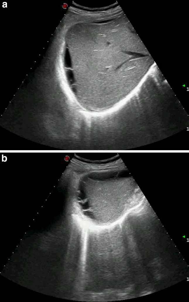
A biloma is an encapsulated collection of bile located in the abdomen. It occurs spontaneously or secondary to traumatic or iatrogenic injury to the biliary system. The patient's medical history, symptoms and diagnostic imaging findings suggest the diagnosis, but a definitive diagnosis is provided by drainage and biochemical analysis of the fluid. We report a case of a patient admitted with acute abdominal pain in the right hypochondrium caused by a spontaneous biloma. This is a rare condition, and the reason for the onset was not identified. We discuss the role of the various diagnostic imaging techniques, particularly that of ultrasound.
Il biloma è una raccolta circoscritta di bile in addome, spontanea o secondaria ad una lesione traumatica o iatrogena del sistema biliare. L’anamnesi, i sintomi ed i reperti radiologici suggeriscono la diagnosi, anche se spesso la diagnosi finale è legata al drenaggio del liquido. Riportiamo il caso di un paziente ricoverato per dolore addominale acuto in ipocondrio destro determinato da un biloma spontaneo, condizione rara in cui non si identifica la causa del danno al sistema biliare e discutiamo il ruolo delle varie metodiche di diagnostica per immagine, in particolare dell’ecografia.
Keywords: Contrast-enhanced CT; Imaging; Spontaneous biloma; Ultrasound.
Figures


References
-
- Whipple C. A case of traumatic rupture of the liver: formation of cystic swelling containing bile stained fluid. Lancet. 1898;1:719. doi: 10.1016/S0140-6736(01)87273-1. - DOI
LinkOut - more resources
Full Text Sources
Other Literature Sources
