

Galectin-1: a biomarker of surgical stress in murine model of cardiac surgery
- PMID: 26261609
- PMCID: PMC4525943
Galectin-1: a biomarker of surgical stress in murine model of cardiac surgery
Abstract
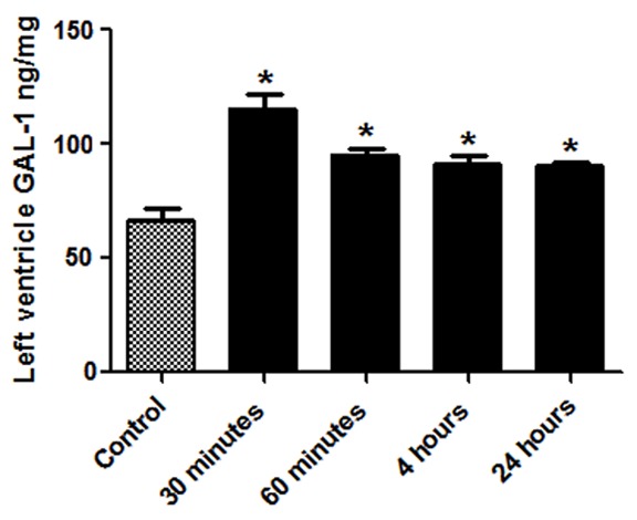
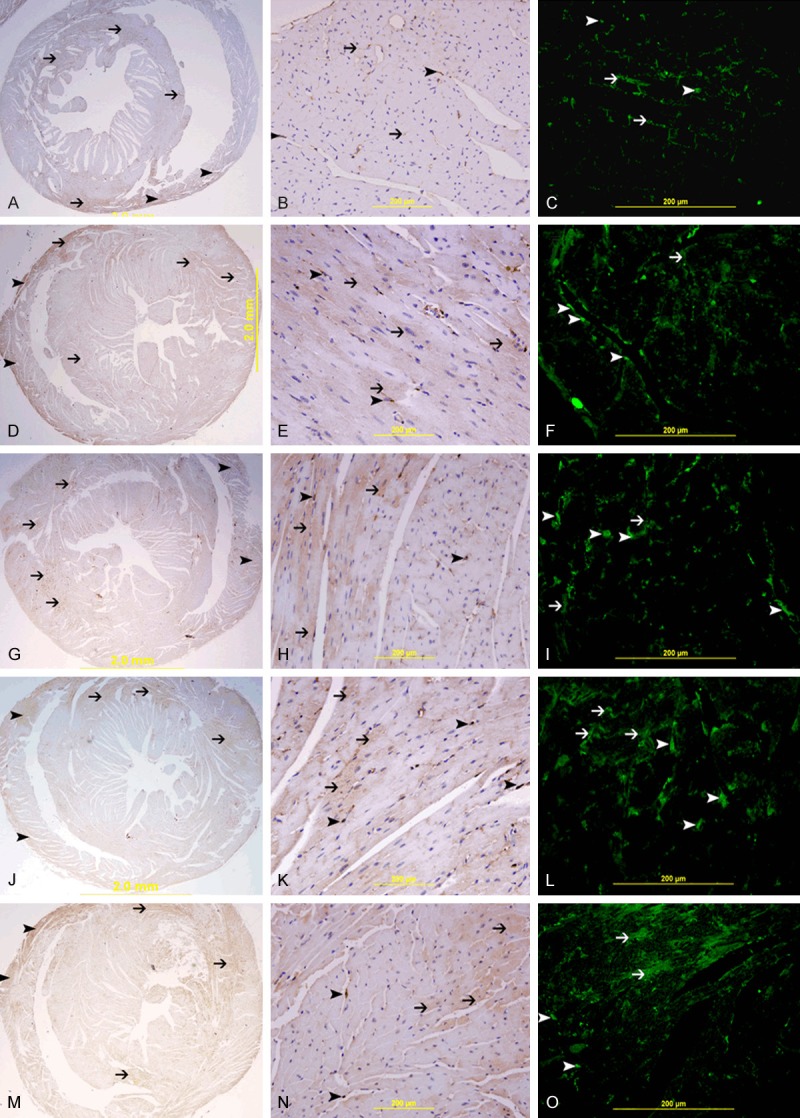
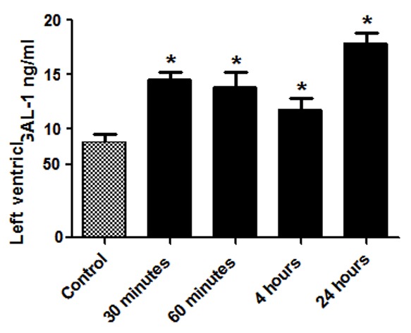
Galectin-1 (GAL-1) belongs to the family of β-galactoside-binding lectins. It regulates cell-cell and cell-matrix interactions, the immune response, apoptosis, cell cycle, RNA splicing and neoplastic transformation. We investigate the effect of heart manipulation secondary to cardiac surgery on the level of GAL-1 in murine heart and plasma. Male C57B6/J mice were used for adopted model of cardiac surgery. Heart samples were processed for immunohistochemical and immunofluorescent labeling, Enzyme linked immunosorbent assay and quantitative RT-PCR to identify GAL-1 levels in the heart and plasma during the first 24 hours following cardiac surgery. There is significant increase of GAL-1 in the LV at 30 minutes (P<0.000), 60 minutes (P<0.001), 4 hour (P<0.003), and 24 hour (P<0.003) time points of surgically operated groups compared to non-operated control group, while GAL-1 mRNA levels in any of the surgically operated groups are not significantly different from the non-operated group suggesting extracardiac origin of this raise of GAL-1. There is significant increase of GAL-1 in the plasma at 30 minutes (P<0.000), 60 minutes (P<0.009), 4 hour (P<0.043), and 24 hour (P<0.000) time points of surgically operated groups compared to non-operated control group. In conclusion, GAL-1 is valuable biomarker of surgical stress.
Keywords: Heart; galectin-1; surgical stress.
Figures
Similar articles
-
Galectin-3 is expressed in the myocardium very early post-myocardial infarction.Cardiovasc Pathol. 2015 Jul-Aug;24(4):213-23. doi: 10.1016/j.carpath.2014.12.001. Epub 2014 Dec 9. Cardiovasc Pathol. 2015. PMID: 25547609
-
Galectin-1 in early acute myocardial infarction.PLoS One. 2014 Jan 31;9(1):e86994. doi: 10.1371/journal.pone.0086994. eCollection 2014. PLoS One. 2014. PMID: 24498007 Free PMC article.
-
Galectin-3 Mitigates Cardiomyocytes Injury through Modulation of Left Ventricular Cathepsins B, D, L and S at 24-Hour Post Myocardial Infarction.Cell Physiol Biochem. 2022 Apr 13;56(2):150-165. doi: 10.33594/000000510. Cell Physiol Biochem. 2022. PMID: 35413750
-
Galectin-1 and galectin-3: plausible tumour markers for oral squamous cell carcinoma and suitable targets for screening high-risk population.Clin Chim Acta. 2015 Mar 10;442:13-21. doi: 10.1016/j.cca.2014.12.038. Epub 2015 Jan 9. Clin Chim Acta. 2015. PMID: 25578395
-
The Galectin-1 level in serum as a novel marker for stress.Glycoconj J. 2010 May;27(4):419-25. doi: 10.1007/s10719-010-9288-z. Glycoconj J. 2010. PMID: 20390448
Cited by
-
Functional Vascular Tissue Engineering Inspired by Matricellular Proteins.Front Cardiovasc Med. 2019 May 31;6:74. doi: 10.3389/fcvm.2019.00074. eCollection 2019. Front Cardiovasc Med. 2019. PMID: 31214600 Free PMC article. Review.
References
-
- Barondes SH, Cooper DN, Gitt MA, Leffler H. Galectins. Structure and function of a large family of animal lectins. J Biol Chem. 1994;269:20807–10. - PubMed
-
- Poirier F. Roles of galectins in vivo. Biochem Soc Symp. 2002:95–103. - PubMed
-
- Poirier F, Timmons PM, Chan CT, Guénet JL, Rigby PW. Expression of the L14 lectin during mouse embryogenesis suggests multiple roles during pre- and post-implantation development. Development. 1992;115:143–55. - PubMed
-
- Poirier F, Robertson EJ. Normal development of mice carrying a null mutation in the gene encoding the L14 S-type lectin. Development. 1993;119:1229–36. - PubMed
Publication types
MeSH terms
Substances
LinkOut - more resources
Full Text Sources
Medical
Research Materials