

Diagnosis and management of cystic lesions of the pancreas
- PMID: 26261724
- PMCID: PMC4502158
- DOI: 10.3978/j.issn.2078-6891.2015.057
Diagnosis and management of cystic lesions of the pancreas
Abstract
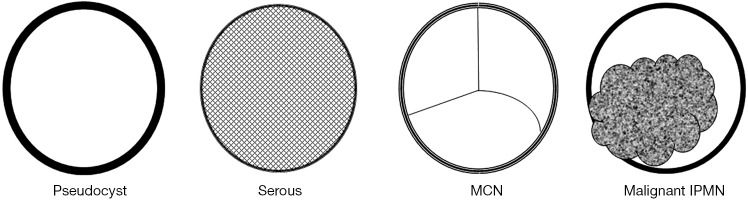
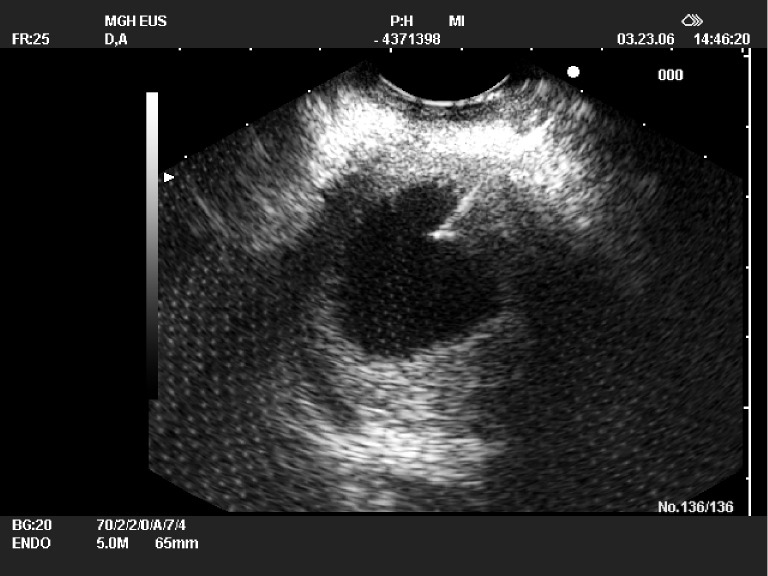
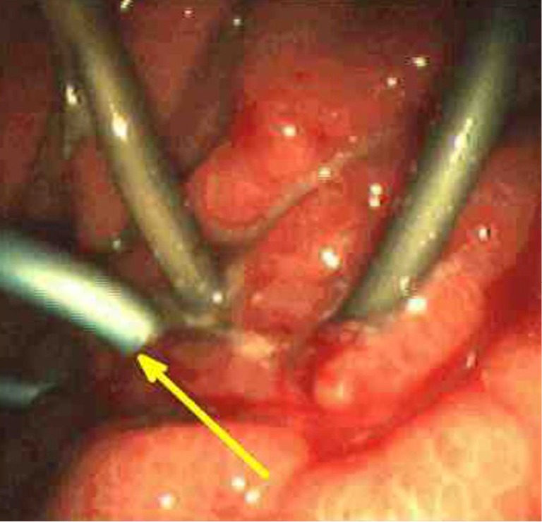
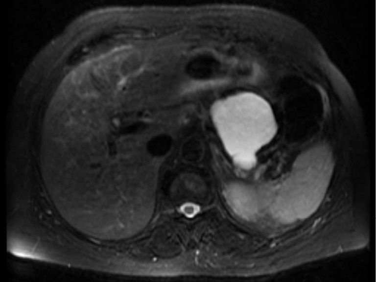
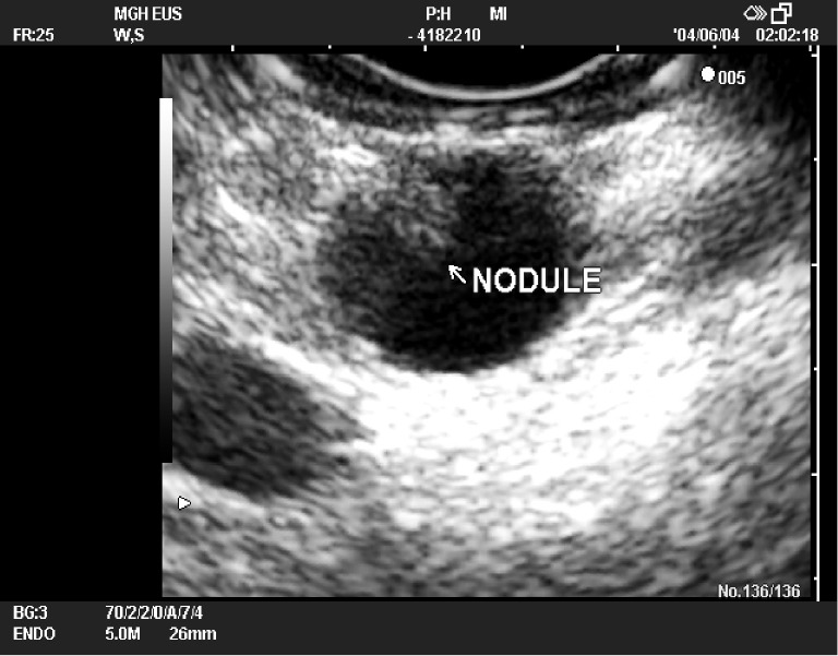
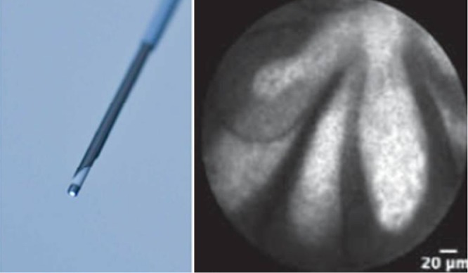
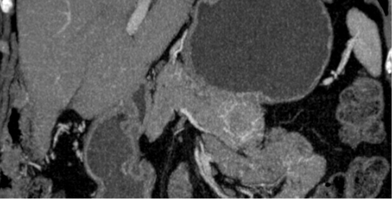
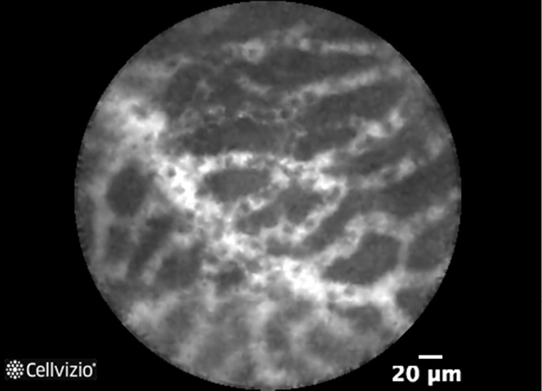
Pancreatic cystic lesions (PCLs) are being increasingly identified in recent years. They show a wide spectrum of imaging and clinical features. The diagnosis and discrimination of these lesions are very important because of the risk for concurrent or later development of malignancy. PCLs are usually first diagnosed and characterized by conventional imaging modalities such as trans-abdominal ultrasonography (US), computed tomography (CT) and magnetic resonance imaging (MRI). However, their ability to differentiate the benign and malignant lesions remains limited. Endoscopic US may be more helpful for the diagnosis and differentiation of PCLs because of its high resolution and better imaging characteristics than cross-sectional imaging modalities. It also allows for fine-needle aspiration (FNA) of cystic lesions for biochemical, cytological and DNA analysis that might be further helpful for diagnosis and differentiation. The management options of PCLs are to observe, endoscopic treatment or surgical resection. However, the decision for management is sometimes hampered by limitations in current diagnostic and tissue sampling techniques. As further diagnostic and non-invasive management options become available, clinical decision-making will become much easier for these lesions.
Keywords: Pancreas; cystic lesions; endoscopic ultrasonography (endoscopic US); intraductal papillary mucinous neoplasms (IPMNs); mucinous cyst; pseudocyst.
Figures

References
-
- Yoon WJ, Brugge WR. Pancreatic cystic neoplasms: diagnosis and management. Gastroenterol Clin North Am 2012;41:103-18. - PubMed
-
- Moparty B, Brugge WR. Approach to pancreatic cystic lesions. Curr Gastroenterol Rep 2007;9:130-5. - PubMed
-
- Kimura W, Nagai H, Kuroda A, et al. Analysis of small cystic lesions of the pancreas. Int J Pancreatol 1995;18:197-206. - PubMed
Publication types
LinkOut - more resources
Full Text Sources
Other Literature Sources
Research Materials