. 2015 Aug 11;10(8):e0135217.
doi: 10.1371/journal.pone.0135217. eCollection 2015.

Efficient Generation of Plasmacytoid Dendritic Cell from Common Lymphoid Progenitors by Flt3 Ligand

Affiliations

Efficient Generation of Plasmacytoid Dendritic Cell from Common Lymphoid Progenitors by Flt3 Ligand

Yi-Ling Chen et al. PLoS One. .

Abstract

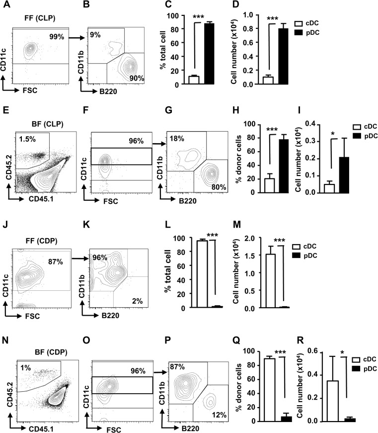
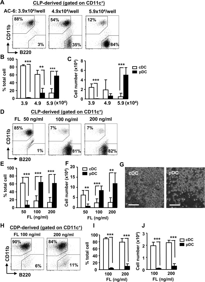
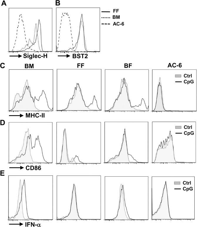
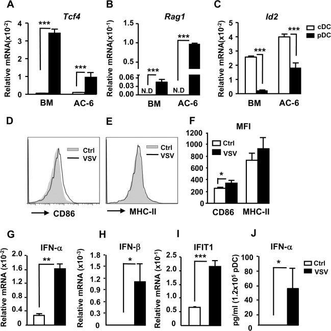
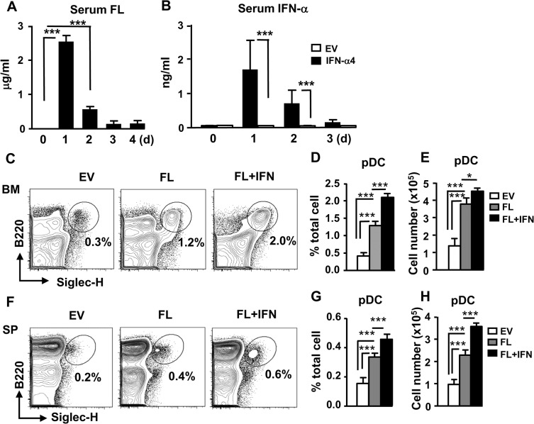
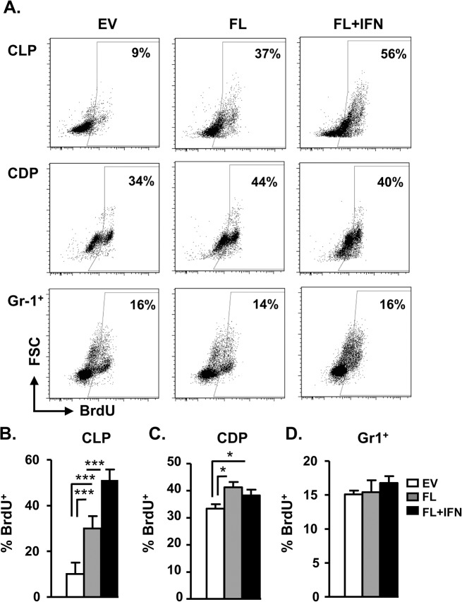
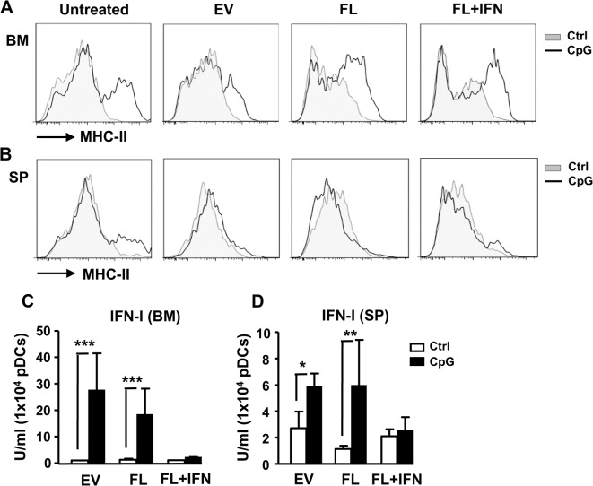
Dendritic cells (DCs), including conventional DCs (cDCs) and plasmacytoid DCs (pDCs) are critical for initiating and controlling the immune response. However, study of DC, particularly pDC, function is hampered by their low frequency in lymphoid organs, and existing methods for in vitro DC generation preferentially favor the production of cDCs over pDCs. Here, we demonstrated that pDCs could be efficiently generated in vitro from common lymphoid progenitors (CLPs) using Flt3 ligand (FL) in three different culture systems, namely feeder-free, BM-feeder and AC-6-feeder. This was in stark contrast to common DC progenitors (CDPs), in which cDCs were prominently generated under the same conditions. Moreover, the efficiency and function of pDCs generated from these three systems varied. While AC-6 system showed the greatest ability to support pDC development from CLPs, BM-feeder system was able to develop pDCs with better functionality. pDCs could also be expanded in vivo using hydrodynamic gene transfer of FL, which was further enhanced by the combined treatment of FL and IFN-α. Interestingly, IFN-α selectively promoted the proliferation of CLPs and not CDPs, which might contribute to enhanced pDC development. Together, we have defined conditions for in vitro and in vivo generation of pDCs, which may be useful for investigating the biology of pDCs.


Conflict of interest statement

Competing Interests: The authors have declared that no competing interests exist.

Figures

Fig 1
Fig 1. In vitro development of pDCs from CLPs.
(A-D) CD45.2+ CLPs (1x10⁴) were cultured alone (feeder-free or FF, n = 3) or (E-I) cocultured with CD45.1xCD45.2 (2x10⁵) BM cells (BM-feeder or BF, n = 3–5) in the presence of 100 ng/ml FL for 6 d. (J-M) Same as in (A-I), except CD45.2+ CDPs were used. The progeny cells were stained with antibodies to CD11c, CD11b, and B220 and analyzed for cDCs (CD11c+CD11b+B220-) and pDCs (CD11c+CD11b-B220+) by flow cytometry. Mean percentages and cell numbers of cDCs and pDCs are shown. *, p<0.05, ***, p<0.005.
Fig 2
Fig 2. Dose-dependent effects of AC-6 feeder cells or FL on DC development from CLPs.
(A) Sorted CLPs (5x10² cells) were cocultured with the indicated numbers of AC-6 in the presence of FL (100 ng/ml) for 20 d. The progeny cells were stained with antibodies to CD11c, CD11b, and B220, gated on CD11c+ and analyzed for cDCs (CD11c+CD11b+B220-) and pDCs (CD11c+CD11b-B220+) by flow cytometry. Mean percentages (B) and cell numbers (C) of cDCs and pDCs are shown (n = 3–7). (D) Same as in (A), except CLPs were cocultured with 5.9x10⁴ AC-6 in the presence of the indicated doses of FL. Mean percentages (E) and cell numbers (F) of cDCs and pDCs are shown (n = 5–15). (G) Images of light microscopy of AC-6-derived cDC and pDC colonies are shown (scale bar 50 μm). (H) Same as in (D-F), except CDPs were cocultured with 5.9x10⁴ AC-6 in the presence of the indicated doses of FL for 12 d. Mean percentages (I) and cell numbers (J) of cDCs and pDCs are shown (n = 3). **, p<0.01, ***, p<0.005.
Fig 3
Fig 3. Maturation and function of in vitro generated pDCs.
Surface expressions of Siglec-H (A) and BST2 (B) on pDCs derived from CLPs using FF, BF and AC-6 systems were shown (n = 3). (C) pDCs sorted from BM or BF culture system or DCs derived from FF and AC-6 culture system were treated with or without CpG ODN (1 μg/ml) for 24 h. Surface staining of MHC class II (C) and CD86 (D) or intracellular staining of IFN-α (E) on sorted BM pDCs or BF-pDCs or gated pDCs from FF- or AC-6 system are shown. One representative experiment out of three.
Fig 4
Fig 4. AC-6-pDCs highly express Tcf4 and Rag1 and produce IFN-I in response to viral infection.
Total RNA prepared from sorted cDCs and pDCs from BM or AC-6 culture system were subjected to RT-qPCR using primers to Tcf4 (A), Rag1 (B) and Id2 (C). Relative mRNA was normalized to Actb. Purified AC-6-pDCs (1.2x10⁵) were infected with or without EMCV or VSV at an MOI of 10 for 18h. The infected cells were stained with antibodies to CD11c, CD11b, B220, MHC class II and CD86 and analyzed by flow cytometry. Expression of CD86 (D) or MHC class II (E) on pDCs (CD11c+CD11b-B220+) is shown. (F) MFI of CD86 and MHC class II on AC-6-pDCs infected with or without VSV was shown. (n = 3) (G-J) Same as in (D-E), except the mRNA of the virus infected pDCs were prepared and subjected to RT-qPCR using primers to all Ifnα (G), Ifnb (H), Ifit1 (I) and Rpl7. Relative mRNA was normalized to Rpl7, a reference gene. (J) Supernatant of the cells infected with virus was subjected to ELISA for IFN-α production. *, p<0.05, **, p<0.01 ***, p<0.005.
Fig 5
Fig 5. IFN-α enhances FL-dependent pDC development in vivo.
Plasmid expressing FL (2.5 μg) or IFN-α4 (0.25 μg) was delivered into WT mice using HGT method. Serum FL (A) and IFN-α (B) was measured by ELISA. (n = 3) (C-H) Same as in (A-B), except empty vector (EV) or plasmid expressing FL (2.5 μg) or FL (2.5 μg) + IFN-α4 (0.25 μg) were delivered in vivo for 6 d. BM (C-E) and spleen cells (F-H) of the treated mice were stained, gated on CD11cintCD11b-, and analyzed for pDCs (CD11cintCD11b-B220+Siglec-H+). Mean percentages and cell numbers of pDCs following the treatments are shown (n = 3). *, p<0.05, ***, p<0.005.
Fig 6
Fig 6. IFN-α enhances FL-dependent proliferation of CLPs, and not CDPs, in vivo.
(A) Empty vector (2.5 μg), plasmid expressing FL (2.5 μg), or FL (2.5 μg) + IFN-α4 (0.25 μg) were delivered into WT mice using HGT method. The mice were then i.p. injected with 0.8 mg of BrdU at last 2 h. Twenty hours post HGT, CLPs (upper) or CDPs (middle) were sorted from the BM and subjected to intracellular staining for BrdU. Similarly, BM was stained and gated on Gr1+ (lower) cells before intracellular staining for BrdU. Mean percentages of BrdU+ cells in CLPs (B), CDPs (C) and Gr1+ cells (D) are shown (n = 3–4). *, p<0.05, ***, p<0.005.
Fig 7
Fig 7. Activation and function of in vivo expanded pDCs.
Empty vector (2.5 μg), plasmid expressing FL (2.5 μg), or FL (2.5 μg) + IFN-α4 (0.25 μg) was delivered in vivo into WT mice using HGT method for 6 d. pDCs sorted from the BM or spleen cells of the treated mice were stimulated with CpG ODN (1 μg/ml) for 24 h. (A-B) The expressions of MHC class II on the sorted BM (A) or spleen (B) pDCs with or without the stimulation were analyzed by flow cytometry. The production of IFN-α by the BM (C) or spleen (D) pDCs following the stimulation was measured by a bioassay as described in the Materials and Methods (n = 3). *, p<0.05, **, p<0.01, ***, p<0.005.

Similar articles

Cited by

References

    1. Gilliet M, Cao W, Liu YJ. Plasmacytoid dendritic cells: sensing nucleic acids in viral infection and autoimmune diseases. Nature reviews Immunology. 2008;8(8):594–606. Epub 2008/07/22. 10.1038/nri2358 . - DOI - PubMed
    1. Reizis B, Bunin A, Ghosh HS, Lewis KL, Sisirak V. Plasmacytoid dendritic cells: recent progress and open questions. Annu Rev Immunol. 2011;29:163–83. Epub 2011/01/12. 10.1146/annurev-immunol-031210-101345 . - DOI - PMC - PubMed
    1. Merad M, Manz MG. Dendritic cell homeostasis. Blood. 2009;113(15):3418–27. Epub 2009/01/30. 10.1182/blood-2008-12-180646 - DOI - PMC - PubMed
    1. Reizis B. Regulation of plasmacytoid dendritic cell development. Curr Opin Immunol. 2010;22(2):206–11. Epub 2010/02/11. 10.1016/j.coi.2010.01.005 - DOI - PMC - PubMed
    1. Shortman K, Sathe P, Vremec D, Naik S, O'Keeffe M. Plasmacytoid dendritic cell development. Adv Immunol. 2013;120:105–26. Epub 2013/09/28. 10.1016/b978-0-12-417028-5.00004-1 . - DOI - PubMed

Publication types

MeSH terms

LinkOut - more resources