. 2015 Aug 11:5:12910.
doi: 10.1038/srep12910.

Human iPSC derived disease model of MERTK-associated retinitis pigmentosa

Affiliations

Human iPSC derived disease model of MERTK-associated retinitis pigmentosa

Dunja Lukovic et al. Sci Rep. .

Abstract

Retinitis pigmentosa (RP) represents a genetically heterogeneous group of retinal dystrophies affecting mainly the rod photoreceptors and in some instances also the retinal pigment epithelium (RPE) cells of the retina. Clinical symptoms and disease progression leading to moderate to severe loss of vision are well established and despite significant progress in the identification of causative genes, the disease pathology remains unclear. Lack of this understanding has so far hindered development of effective therapies. Here we report successful generation of human induced pluripotent stem cells (iPSC) from skin fibroblasts of a patient harboring a novel Ser331Cysfs*5 mutation in the MERTK gene. The patient was diagnosed with an early onset and severe form of autosomal recessive RP (arRP). Upon differentiation of these iPSC towards RPE, patient-specific RPE cells exhibited defective phagocytosis, a characteristic phenotype of MERTK deficiency observed in human patients and animal models. Thus we have created a faithful cellular model of arRP incorporating the human genetic background which will allow us to investigate in detail the disease mechanism, explore screening of a variety of therapeutic compounds/reagents and design either combined cell and gene- based therapies or independent approaches.


Figures

Figure 1
Figure 1
(A). MERTK DNA sequence chromatogram around c.992_993 in healthy and affected patient carrying a deletion of CA (c.992_993delCA). DNA sequencing, performed on healthy and patient’s dermal fibroblasts and iPSCs’ gDNA, confirmed the mutation in early-onset RP patient. (B). The wild type sequence codes for 999 amino acid protein with Immunoglobulin Ig-like C2-type (Ig-like) (residues 100–194 and 203–281), fibronectin type III-like (FN3) (residues 284–368 and 384–470) transmembrane (TM) (residues 502–524) and kinase domain (residues 587–854). The mutation results in a predictive truncated protein of 334 amino acids with expected molecular weight of 36.6 kDa.
Figure 2
Figure 2. Differentiation of healthy and MERTK p.Ser331Cysfs*5 iPSCs into RPE cells.
Bright field micrograph of healthy (A) and MERTK p.Ser331Cysfs*5 (B) iPSC-RPE showing typical RPE morphology, polygonal shape and pigmentation. Semithin sections of iPSC-RPE cells stained with toluidine blue. Both healthy (C) and MERTK p.Ser331Cysfs*5 (D) iPSC-RPE cells form a monolayer of cuboid cells highly polarized with abundant apical microvilli (arrows) and melanosomes (arrowheads). Electron micrograph of cultured iPSC-RPE, healthy (E) and MERTK p.Ser331Cysfs*5 (F,G,H,I). (G) High magnification of melanin granules showing different stages of development. (H) Intercellular junctional complexes which include the tight junctions, adherens junctions (arrows) and membrane interdigitations (arrowheads). (I) The basement membrane of the cells tightly bound to the transwells support film (arrowheads) and small basal infoldings (arrows). A,B scale bar 50µm; C,D scale bar 10µm.
Figure 3
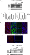
Figure 3. RPE characteristic marker gene and protein expression in iPSC-RPE from healthy individual and RP patient.
(A. i) BEST1, CRALBP, RPE65, MERTK gene expression in fibroblasts, iPSCs and iPSC-RPE. Two iPSC lines (iPSCc1 and iPSCc2) and their respective iPSC-RPE from each individual are analyzed. The samples were loaded on agarose gels prepared and run under the same experimental conditions. (ii) Quantitative reverse transcription polymerase chain reaction to analyze expression of genes characteristic for RPE. Fold expression in undifferentiated iPSCs and iPSC-RPE from healthy and MERTK p.Ser331Cysfs*5 patient normalized to their originating dermal fibroblasts (DF). Each bar represents the average ±SEM of at least three independent biological replicates (* p ≤ 0.05, **p ≤ 0.05, ***p ≤ 0.05). (B. i) Expression of RPE characteristic proteins by iPSC-RPE. Immunocytochemistry against RPE65, CRALBP, BEST1, ZO-1 in healthy and MERTK p.Ser331Cysfs*5 iPSC-RPE. Images were taken with Leica confocal microscope TCS SP5 using HCX PL APO lambda blue 63X/ 1.4 OIL objective. Scale bar 10µm. (ii) MERTK expression in iPSC-RPE. Immunocytochemistry against MERTK. Apical section and vertical section simulation showing apical MERTK distribution in the healthy individual. MERTK p.Ser331Cysfs*5 iPSC-RPE stain negative for MERTK at all sections. Images were taken with Leica confocal microscope TCS SP5 using HCX PL APO lambda blue 63X/ 1.4 OIL objective, scale bar 10 μm. (C). Western blot analyses of RPE-specific marker protein expression in fibroblasts (DF), two iPSC lines (iPSCc1 and iPSCc2) and their respective iPSC-RPE from each individual are analyzed. The expression of CRALBP and BEST1 is detected only in iPSC- RPE and hRPE cells. β actin was used as loading control. Cropped blots are from gels run under the same experimental conditions and loaded with the same samples.
Figure 4
Figure 4. In vitro phagocytosis assay of photoreceptor outer segments (OS) by iPS-RPE from healthy individual and MERTK p.Ser331Cysfs*5 patient.
Basolateral sections across the iPSC-RPE cells together with vertical section simulation. Healthy iPS-RPE internalize FITC-labeled OS (green) while patient’s one do not. F-actin is stained by phalloidin (red) to visualize cell morphology. Images were taken with Leica confocal microscope TCS SP8 using HCX PL APO lambda blue 63X/ 1.4 OIL objective, scale bar 25 μm.

References

    1. Hartong D. T., Berson E. L. & Dryja T. P. Retinitis pigmentosa. Lancet 368, 1795–1809, 10.1016/S0140-6736(06)69740-7 (2006). - DOI - PubMed
    1. Takahashi K. et al. Induction of pluripotent stem cells from adult human fibroblasts by defined factors. Cell 131, 861–872, 10.1016/j.cell.2007.11.019 (2007). - DOI - PubMed
    1. Lamba D. A. et al. Generation, purification and transplantation of photoreceptors derived from human induced pluripotent stem cells. PloS one 5, e8763, 10.1371/journal.pone.0008763 (2010). - DOI - PMC - PubMed
    1. Ikeda H. et al. Generation of Rx+/Pax6+ neural retinal precursors from embryonic stem cells. Proceedings of the National Academy of Sciences of the United States of America 102, 11331–11336, 10.1073/pnas.0500010102 (2005). - DOI - PMC - PubMed
    1. Osakada F., Ikeda H., Sasai Y. & Takahashi M. Stepwise differentiation of pluripotent stem cells into retinal cells. Nature protocols 4, 811–824, 10.1038/nprot.2009.51 (2009). - DOI - PubMed

Publication types

MeSH terms

Substances