. 2015 Aug 11;10(8):e0135577.
doi: 10.1371/journal.pone.0135577. eCollection 2015.

LTBP-2 Has a Single High-Affinity Binding Site for FGF-2 and Blocks FGF-2-Induced Cell Proliferation

Affiliations

LTBP-2 Has a Single High-Affinity Binding Site for FGF-2 and Blocks FGF-2-Induced Cell Proliferation

Clementine Menz et al. PLoS One. .

Abstract

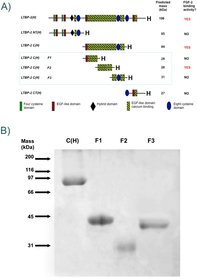
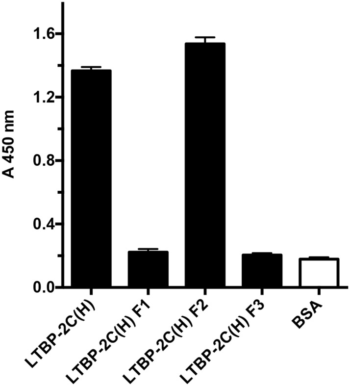
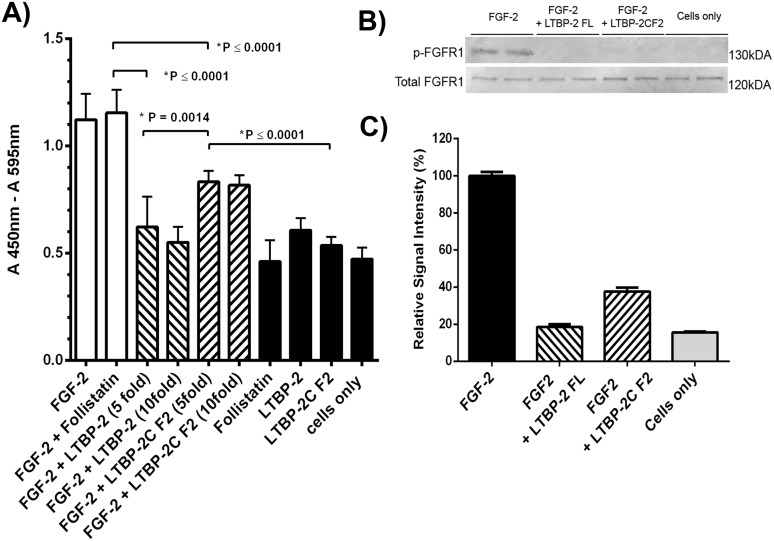
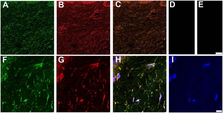
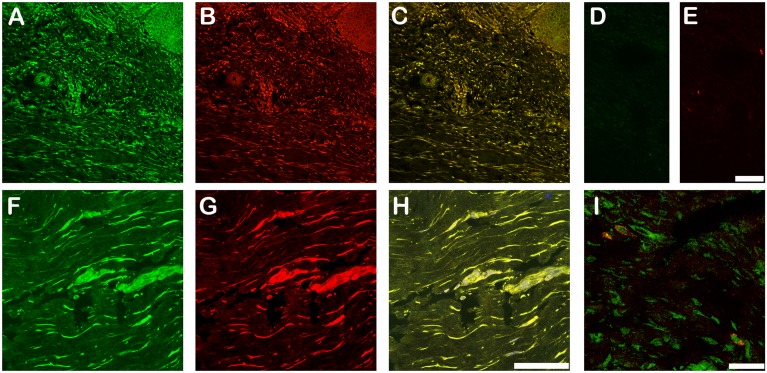
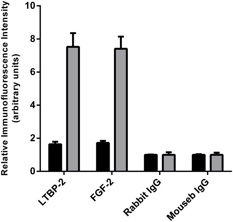
Latent transforming growth factor-beta-1 binding protein-2 (LTBP-2) belongs to the fibrillin-LTBP superfamily of extracellular matrix proteins. LTBPs and fibrillins are involved in the sequestration and storage of latent growth factors, particularly transforming growth factor β (TGF-β), in tissues. Unlike other LTBPs, LTBP-2 does not covalently bind TGF-β, and its molecular functions remain unclear. We are screening LTBP-2 for binding to other growth factors and have found very strong saturable binding to fibroblast growth factor-2 (FGF-2) (Kd = 1.1 nM). Using a series of recombinant LTBP-2 fragments a single binding site for FGF-2 was identified in a central region of LTBP-2 consisting of six tandem epidermal growth factor-like (EGF-like) motifs (EGFs 9-14). This region was also shown to contain a heparin/heparan sulphate-binding site. FGF-2 stimulation of fibroblast proliferation was completely negated by the addition of 5-fold molar excess of LTBP-2 to the assay. Confocal microscopy showed strong co-localisation of LTBP-2 and FGF-2 in fibrotic keloid tissue suggesting that the two proteins may interact in vivo. Overall the study indicates that LTBP-2 is a potent inhibitor of FGF-2 that may influence FGF-2 bioactivity during wound repair particularly in fibrotic tissues.


Conflict of interest statement

Competing Interests: The authors have declared that no competing interests exist.

Figures

Fig 1
Fig 1. Recombinant LTBP-2 Fragments.
A. Schematic diagram of recombinant LTBP-2 fragments. Protein fragments generated specifically for this study (LTBP-2C(H) F1, F2 and F3) are highlighted within the blue box. FGF-2 binding was confined to a single central region of the LTBP-2 molecule consisting of 6 EGF-like repeats (fragment LTBP-2C(H) F2).B. SDS-PAGE of purified recombinant LTBP-2 fragments. Samples of purified fragments LTBP-2 C(H), LTBP-2 C(H) F1, LTBP-2 C(H) F2 and LTBP-2 C(H) F3 were analyzed on a 12% gel under non-reducing conditions and stained with Coomassie blue. The relative mobilities of protein standards are indicated by arrows.
Fig 2
Fig 2. LTBP-2 specifically binds FGF-2 but not VEGF, BMP-4, BMP-7 or TGF-beta.
A. Microtitre wells were coated with rLTBP-2 (black columns) or BSA(shaded columns) (100 ng/ well). After blocking, triplicate wells were incubated at 37°C for 2h with TGF-beta (13 ng / well), VEGF (21 ng / well), BMP-7 (4 ng/well), BMP-4 (4 ng / well) or FGF-2 (10 ng / well). Growth factor binding was detected using specific biotinylated antibodies from Duoset kits as described in material and methods. Mean values ± S.D. from triplicate wells are shown. B. Microtitre wells were coated with rLTBP-2 (100ng/well) was coated onto microtitre plates. After blocking, triplicate wells were incubated at 37°C for 2h with (black columns) or without (cross-hatched) growth factor, (BMP-4 (4ng/well) or FGF-2 (10ng/well). Binding of growth factor to LTBP-2 was detected using biotinylated anti-BMP-4 detection antibody (0.5ug/ml) or anti-FGF-2 detection antibody (0.25ug/ml), followed by a peroxidase detection method (see material and methods). Mean values ± S.D. from triplicate wells are shown. Note the anti-BMP-4 antibody bound to the wells equally strongly in the presence or absence of added BMP-4, indicating the interaction was non-specific.
Fig 3
Fig 3. LTBP-2 interacts strongly with FGF-2.
A. Microtitre wells were coated with 200 ng rLTBP-2 or BSA control. After blocking, triplicate wells were incubated with 0–1.8 nM concentrations of FGF-2 (0–30 ng/ml) for 3 h at 37°C. FGF-2 binding was detected following sequential incubation of the wells with biotinylated mouse anti-[human FGF-2] antibody and streptavidin-HRP conjugate following the duoset protocol. Circles, LTBP-2; squares, BSA. Mean values ± S.D. of triplicate determinations are shown. B. Kd calculation. Following subtraction of the average BSA signal, the A450nm values were converted to fmol of FGF-2 using a standard ELISA curve (not shown). An additional graph was plotted of bound versus added FGF-2 and the Kd for interaction with LTBP-2 was calculated by non-linear regression analysis of the curve using the prism 4.0 program. Mean values ± S.D. from triplicate determinations are shown.
Fig 4
Fig 4. FGF-2 has a single binding domain in the central region of LTBP-2.
A. Three recombinant fragments spanning the LTBP-2 molecule were tested for binding to FGF-2 in a solid phase assay. Full length LTBP-2(H), fragments LTBP-2 NT (H), LTBP-2C (H), LTBP-2 CT (H) or BSA control were coated onto wells at 100 ng/ml, followed by incubation with FGF-2 (100ng/ml) for 3h at 37°C. Strong specific binding to central fragment LTBP-2C(H) was detected as described in Fig 2A. Mean values ± S.D. from triplicate wells are shown. B. A binding curve was produced for the FGF-2 interaction with fragment LTBP-2C(H) following the protocol described under Fig 2, with 400 ng/well (4.8 pmol) of LTBP-2C (H) or BSA control coated on the wells incubated with increasing concentrations FGF-2 (0–1.5 nM). The Kd for binding of FGF-2 to fragment LTBP-2C (H) was calculated as 1.02 ± 0.19 nM. Mean values ± S.D. from triplicate determinations are shown. C. Three sub-fragments F1, F2 and F3 spanning fragment LTBP-2 C(H) were produced and tested for FGF-2 binding as described under Fig 2. LTBP-2C (H) (200 ng/well, 2.4 pmol) or sub-fragment (F1, F2 or F3) (66ng/well, 2.4 pmol) or BSA control was coated on the wells and incubated with FGF-2 (100 ng/ml). Strong specific binding of FGF-2 to sub-fragment LTBP-2C F2 was detected. Mean values ± S.D. from triplicate wells are shown. D. Subsequently binding curves were obtained for sub-fragments F1 (solid squares), F2 (open circles), F3 (solid circles) (35 ng/well, 1.2 pmol) coated on the wells and incubated with increasing concentrations of FGF-2 (0–30 ng / ml). Note specific FGF-2 binding to sub-fragment LTBP-2C F2 but no binding of fragments F1 and F3 above the BSA control (triangles). Mean values ± S.D. from triplicate determinations are shown. E. The Kd for the FGF-2 interaction with sub-fragment LTBP-2C F2 was calculated as 1.03 ± 0.10 nM which is similar to the Kds calculated for the interactions of FGF-2 with full-length LTBP-2 and fragment LTBP2C. Mean values ± S.D. from triplicate determinations are shown.
Fig 5
Fig 5. The FGF-2 binding site is close to the central heparin binding site on LTBP-2.
In a previous study [32] we identified LTBP-2 C(H) as a heparin-binding fragment of LTBP-2. To further define the location of this heparin binding activity, the three sub-fragments F1, F2, F3 spanning LTBP-2 C(H), were assayed for heparin binding using a heparin-albumin conjugate (HAC). HAC or BSA control (400 ng) was coated on wells followed by incubation with equimolar concentrations (23.5 nM) of LTBP-2C(H) or sub-fragment F1, F2 or F3. Specific binding was detected using anti-His4 antibody targeting the poly-His tag on each recombinant fragment. Fragment F2 showed strong specific binding to the heparin conjugate in contrast to F1 and F3 which showed no binding above background. Mean values ± S.D. from triplicate wells are shown.
Fig 6
Fig 6. LTBP-2 blocks FGF-2-induced cell proliferation.
A. The effect of LTBP-2 on the bio-activity of FGF-2 was tested in a cell proliferation assay (see experimental). Human foreskin fibroblasts were treated with FGF-2 with and without follistatin (white columns), or FGF-2 and follistatin pre-incubated with 5 or 10 fold molar excess of full length LTBP-2 or fragment LTBP-2C F2 (cross-hatched). Negative controls (black columns), included cells only and cells incubated with follistatin, LTBP-2 or fragment LTBP-2C F2. Mean values ± S.D. from triplicate determinations. Note 5 fold molar excess of full-length LTBP-2 completely blocked FGF-2 induced cell proliferation (p = 0.0001) and 5-fold molar excess of fragment LTBP-2C F2 partially blocked the activity (p = 0.0001). B. Immunoblot analysis FGF receptor (FGFR1) phosphorylation. Human foreskin fibroblasts were treated for 2 hours with FGF-2 (10 ng / ml) only or with FGF-2 plus 10-fold molar excess of full length LTBP-2 (LTBP-2 FL) or fragment F2 (LTBP-2C F2). Control cells had no FGF-2 or LTBP-2 added. Cellular proteins were extracted and duplicate samples were analysed by SDS-PAGE and immunoblotting with anti-phospho-FGFR1 antibody, and anti-total FGFR1 antibody. Bands were visualised using the LI-COR Odyssey Infrared Imaging System. C. The band intensity was measured using ImageJ 1.48 software [National Institutes of Health (NIH), Bethesda, MD] and normalised to the internal β actin signal. The ratio of the phospho-FGFR1 to total FGFR1 value for each sample is expressed relative to the average FGF-2 only control value (= 100%). Note the strong FGFR1 activation by FGF-2 was substantially blocked by both LTBP-2 C and LTBP-2C F2 fragments. Mean values ± S.D. of duplicate lanes.
Fig 7
Fig 7. LTBP-2 and fibrillin-1 colocalize in fibrotic skin.
Keloid tissue was prepared and analyzed by confocal microscopy as described in the methods section. A and F, polyclonal anti-[human LTBP-2 peptide] antibody 3504 (2 μg/ ml) detected with anti-rabbit IgG antibody conjugated to fluor Alexa 488; B and G, monoclonal anti-[fibrillin-1] antibody MAB1919 (Merck millipore) (2.5 μg/ml) detected with anti-mouse IgG antibody conjugated to Alexa 594; C, A and B merged; D, rabbit IgG control (2 μg/ ml); E, mouse IgG control (2.5 μg / ml); H, F, G and I merged; I, DAPI nuclear stain. Magnification: top row, Bar = 100 μm; bottom row, Bar = 10 μm.
Fig 8
Fig 8. LTBP-2 and FGF-2 co-localize in keloid tissue.
Keloid tissue was also analyzed for LTBP-2 and FGF-2 by confocal microscopy. A and F, polyclonal anti-[human LTBP-2 peptide] antibody 3504 (2 μg/ ml) detected with anti-rabbit IgG antibody conjugated to fluor Alexa 488; B and G, monoclonal anti-[human FGF-2] antibody #61087 (BD Biosciences) (2.5 μg/ml) detected with anti-mouse IgG antibody conjugated to Alexa 594; C, A and B merged; D, rabbit IgG control (2 μg/ ml); E, mouse IgG control (2.5 μg / ml); H, F, and G merged; I, Control confocal image showing distinct immunostaining patterns for VEGF (red) and LTBP-2 (green). Magnification: top row, Bar = 100 μm; bottom row, Bar = 50 μm.
Fig 9
Fig 9. Quantitation of LTBP-2 and FGF-2 in normal skin and keloid.
The relative fluorescence intensities of LTBP-2 and FGF-2 staining (and appropriate IgG controls) in sections of normal human skin (black columns) and keloid (shaded columns) was quantitated from 3 random areas (each 0.038 mm2) per section using the AnalySIS software package (Soft-Imaging System, Munster, Germany). Values expressed relative to the background control signal (= 1 unit). Mean values ± S.D. of triplicate determination are shown.

References

    1. Hyytiainen M, Penttinen C, Keski-Oja J (2004) Latent TGF-beta binding proteins: extracellular matrix association and roles in TGF-beta activation. Crit Rev Clin Lab Sci 41: 233–264. - PubMed
    1. Kielty CM, Sherratt MJ, Marson A, Baldock C (2005) Fibrillin microfibrils. Adv Protein Chem 70: 405–436. - PubMed
    1. Rifkin DB (2005) Latent transforming growth factor-beta (TGF-beta) binding proteins: orchestrators of TGF-beta availability. J Biol Chem 280: 7409–7412. - PubMed
    1. Baldwin AK, Simpson A, Steer R, Cain SA, Kielty CM (2013) Elastic fibres in health and disease. Expert Rev Mol Med 15: e8 10.1017/erm.2013.9 - DOI - PubMed
    1. Ramirez F, Sakai LY (2010) Biogenesis and function of fibrillin assemblies. Cell Tissue Res 339: 71–82. 10.1007/s00441-009-0822-x - DOI - PMC - PubMed

Publication types

MeSH terms

LinkOut - more resources