Case Reports
. 2015 May;97(4):e64-6.
doi: 10.1308/003588415X14181254789682.

Radiological considerations and surgical planning in the treatment of giant parathyroid adenomas

Affiliations
Case Reports

Radiological considerations and surgical planning in the treatment of giant parathyroid adenomas

G Garas et al. Ann R Coll Surg Engl. 2015 May.

Abstract

Giant parathyroid adenomas constitute a rare clinical entity, particularly in the developed world. We report the case of a 53-year-old woman where the initial ultrasonography significantly underestimated the size of the lesion. The subsequent size and weight of the adenoma (7 cm diameter, 27 g) combined with the severity of the hypercalcaemia raised the suspicion for the presence of a parathyroid carcinoma. This was later disproven by the surgical and histological findings. Giant parathyroid adenomas are encountered infrequently among patients with primary hyperparathyroidism, and appear to have distinct clinical and biochemical features related to specific genomic alterations. Cross-sectional imaging is mandated in the investigation of parathyroid adenomas presenting with severe hypercalcaemia as ultrasonography alone can underestimate their size and extent. This is important since it can impact on preoperative preparation and planning as well as the consent process as a thoracic approach may prove necessary for certain cases.

Keywords: Consent; Giant; Imaging; Parathyroid adenoma; Primary hyperparathyroidism; Surgery.


Figures

Figure 1
Figure 1
Ultrasonography demonstrating a lobular (arrows indicating lobules), well defined hypoechoic lesion situated behind the left lower pole of the thyroid gland
Figure 2
Figure 2
T2 weighted coronal magnetic resonance imaging illustrating a left inferior parathyroid adenoma (arrow) extending deep into the mediastinum down to the origin of the left common carotid artery
Figure 3
Figure 3
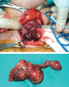
Intraoperative (arrow) and immediately postoperative photographs revealing a well defined lobular mass
Figure 4
Figure 4
Histological features of the giant parathyroid adenoma (haematoxylin and eosin stain, 100x magnification): The lesion consists of chief cells (arrow), among which there are many delicate vessels (A). In some areas, nodules of water clear cells (arrow) are present (B).

References

    1. Fraser WD. Hyperparathyroidism. Lancet 2009; 374: 145–158. - PubMed
    1. Lalanne-Mistrih ML, Ognois-Ausse P, Goudet P, Cougard P. Giant parathyroid tumors: characterization of 26 glands weighing more than 3.5 grams. Ann Chir 2002; 127: 198–202. - PubMed
    1. Spanheimer PM, Stoltze AJ, Howe JR et al. Do giant parathyroid adenomas represent a distinct clinical entity? Surgery 2013; 154: 714–718. - PMC - PubMed
    1. Sulaiman L, Nilsson IL, Juhlin CC et al. Genetic characterization of large parathyroid adenomas. Endocr Relat Cancer 2012; 19: 389–407. - PMC - PubMed
    1. Sisodiya R, Kumar S, Palankar N, Bv D. Case report on giant parathyroid adenoma with review of literature. Indian J Surg 2013; 75(Suppl 1): 21–22. - PMC - PubMed

Publication types