

Palatal and retropharyngeal injury secondary to intubation using the GlideScope® video laryngoscope
- PMID: 26263957
- PMCID: PMC4473888
- DOI: 10.1308/003588415X14181254789727
Palatal and retropharyngeal injury secondary to intubation using the GlideScope® video laryngoscope
Abstract
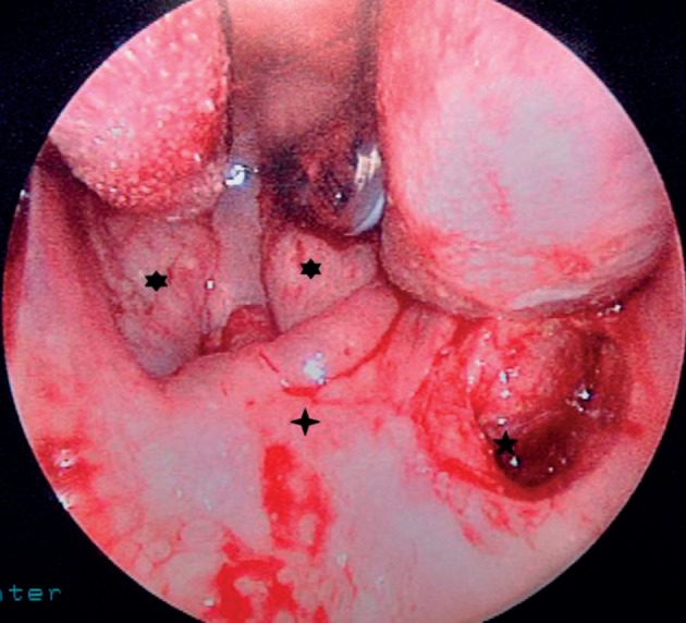
Introduction: There are few reports of injury to the soft palate and retropharynx sustained during intubation with the GlideScope® video laryngoscope. Most reports are of isolated injury to the soft palate.
Case history: We describe a patient in whom the retropharynx was injured but the extent of the injury was not observed initially. The patient did not suffer severe sequelae from this injury. However, this injury can cause serious sequelae if it is not recognised (eg development of a retropharyngeal abscess).
Conclusions: We recommend that any patient who sustains injury to the soft palate during intubation (particularly if the endotracheal tube passes through the soft palate) should be reviewed an otolaryngologist before removal of the endotracheal tube.
Keywords: Endotracheal tube; GlideScope® video laryngoscope; Intubation – Palate.
Figures


References
-
- Cooper RM. Complications associated with the use of the GlideScope video laryngoscope. Can J Anaesth 2007; 54: 54–57. - PubMed
-
- Cross P, Cytryn J, Cheng KK. Perforation of the soft palate using the GlideScope video laryngoscope. Can J Anaesth 2007; 54: 588–589. - PubMed
-
- Leong WL, Lim Y, Sia AT. Palatopharyngeal wall perforation during Glidescope intubation. Anaesth Intens Care 2008; 36: 870–874. - PubMed
-
- Malik AM, Frogel JK. Anterior tonsillar pillar perforation during GlideScope video laryngoscopy. Anesth Analg 2007; 104: 1,610–1,611. - PubMed
-
- Vincent RD Jr, Wimberly MP, Brockwell RC, Magnuson JS. Soft palate perforation during orotracheal intubation facilitated by the GlideScope video laryngoscope. J Clin Anesth 2007; 19: 619–621. - PubMed
Publication types
MeSH terms
LinkOut - more resources
Full Text Sources
Other Literature Sources
Medical