. 2015 Oct;138(Pt 10):2907-19.
doi: 10.1093/brain/awv233. Epub 2015 Aug 10.

Structural imaging biomarkers of sudden unexpected death in epilepsy

Affiliations

Structural imaging biomarkers of sudden unexpected death in epilepsy

Britta Wandschneider et al. Brain. 2015 Oct.

Abstract

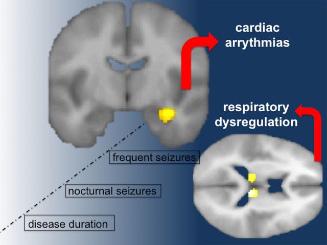
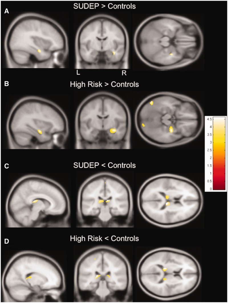
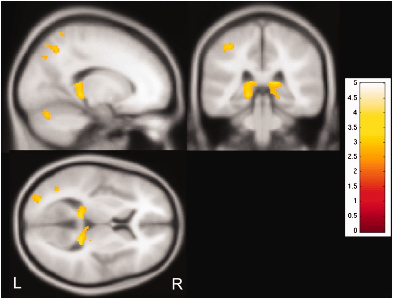
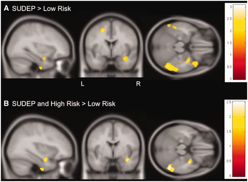
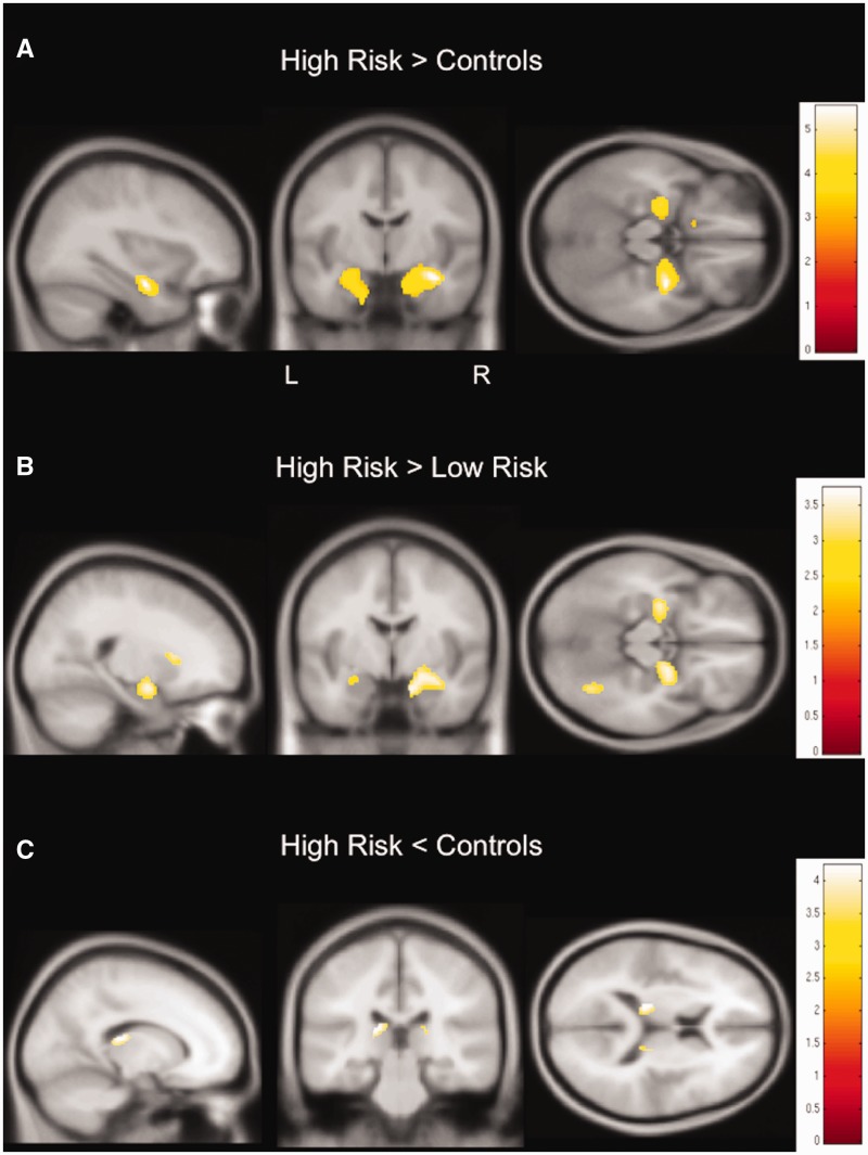
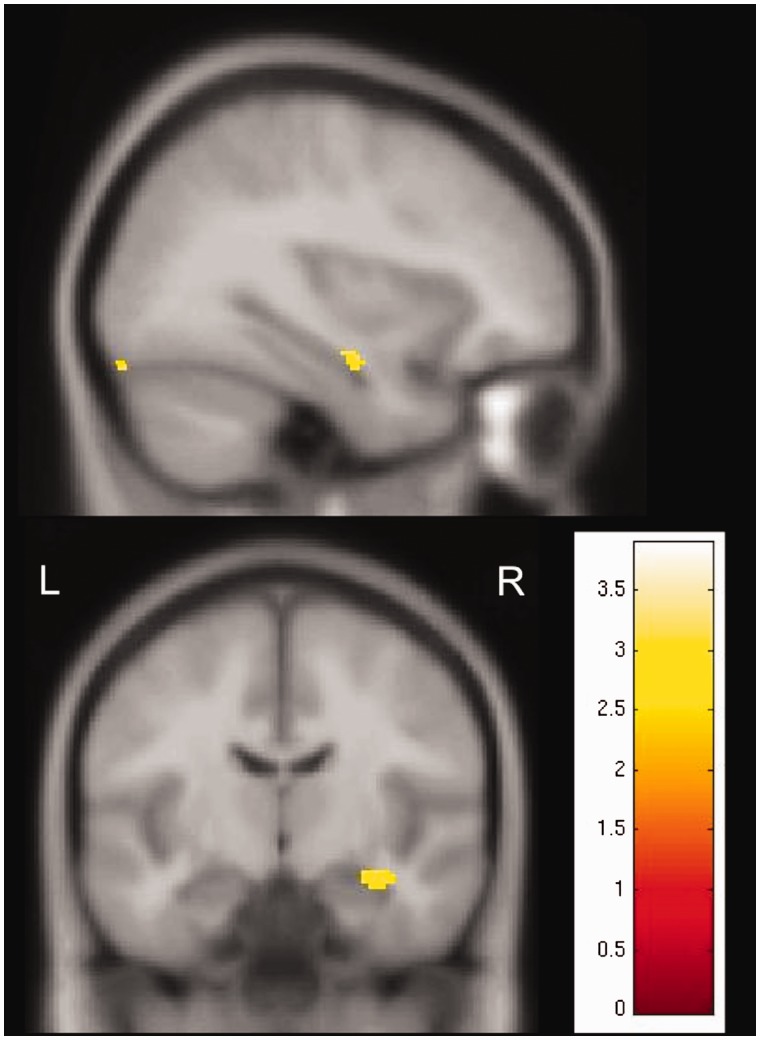
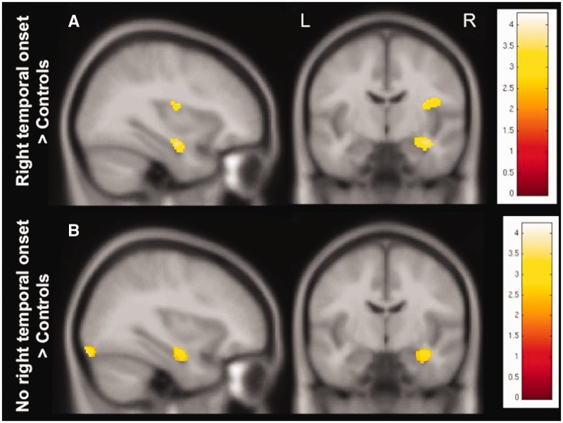
Sudden unexpected death in epilepsy is a major cause of premature death in people with epilepsy. We aimed to assess whether structural changes potentially attributable to sudden death pathogenesis were present on magnetic resonance imaging in people who subsequently died of sudden unexpected death in epilepsy. In a retrospective, voxel-based analysis of T1 volume scans, we compared grey matter volumes in 12 cases of sudden unexpected death in epilepsy (two definite, 10 probable; eight males), acquired 2 years [median, interquartile range (IQR) 2.8] before death [median (IQR) age at scanning 33.5 (22) years], with 34 people at high risk [age 30.5 (12); 19 males], 19 at low risk [age 30 (7.5); 12 males] of sudden death, and 15 healthy controls [age 37 (16); seven males]. At-risk subjects were defined based on risk factors of sudden unexpected death in epilepsy identified in a recent combined risk factor analysis. We identified increased grey matter volume in the right anterior hippocampus/amygdala and parahippocampus in sudden death cases and people at high risk, when compared to those at low risk and controls. Compared to controls, posterior thalamic grey matter volume, an area mediating oxygen regulation, was reduced in cases of sudden unexpected death in epilepsy and subjects at high risk. The extent of reduction correlated with disease duration in all subjects with epilepsy. Increased amygdalo-hippocampal grey matter volume with right-sided changes is consistent with histo-pathological findings reported in sudden infant death syndrome. We speculate that the right-sided predominance reflects asymmetric central influences on autonomic outflow, contributing to cardiac arrhythmia. Pulvinar damage may impair hypoxia regulation. The imaging findings in sudden unexpected death in epilepsy and people at high risk may be useful as a biomarker for risk-stratification in future studies.

Keywords: autonomic; hippocampus; sudden death; sudep risk; voxel based morphometry.


Figures

None
The mechanisms underlying sudden unexpected death in epilepsy (SUDEP) remain unclear. Wandschneider et al. reveal increased amygdalo-hippocampal volume in cases of SUDEP and in individuals at high risk, compared to individuals at low risk and people without epilepsy. Findings are consistent with histopathological reports in sudden infant death syndrome.
Figure 1
Figure 1
Regional grey matter volume differences between SUDEP and people at high risk and controls. (A) SUDEP cases show increased grey matter volume in the right hippocampus and parahippocampal gyrus compared to healthy subjects. (B) Similarly to SUDEP cases, subjects at high risk show increased grey matter volume in the right hippocampus and parahippocampal gyrus compared to healthy controls. (C) Compared to controls, grey matter volume is decreased in SUDEP cases in the pulvinar bilaterally. (D) Likewise, grey matter volume is decreased in those at high risk in the left pulvinar, compared to healthy controls. T-values are represented in the coloured bars. P < 0.001, 30 voxel threshold extent; L = left; R = right.
Figure 2
Figure 2
Correlation of grey matter volume with disease duration. Regional grey matter volume in bilateral thalamic pulvinar shows a negative correlation with epilepsy duration, i.e. grey matter volume decreases with longer duration (P < 0.005, 30 voxel threshold extent). T-values are represented in the coloured bar. L = left; R = right.
Figure 3
Figure 3
Regional grey matter volume differences between SUDEP cases and those at high risk in comparison to people at low risk. (A) Similar to findings in comparison to controls (Fig. 1A), but at a lower threshold level (P < 0.05, 30 voxels threshold extent), SUDEP cases show increased grey matter volume in the right hippocampus and parahippocampal gyrus in comparison to those at low risk. (B) People at high risk and SUDEP cases share common areas of increased grey matter volume within the right hippocampus and parahippocampal gyrus when compared to those at low risk (conjunction, P < 0.05, 30 voxels threshold extent). T-values are represented in the coloured bars. L = left; R = right.
Figure 4
Figure 4
Regional grey matter volume differences between those at low and high risk with non-lesional epilepsy and controls. Findings appear similar to previous findings in the whole sample (Fig. 1), but are more bilateral: subjects at high risk without identifiable pathology on clinical structural scans show an increase of grey matter volume in both anterior hippocampi and amygdalae when compared to controls (A; P < 0.001, 30 voxels threshold extent) and when compared to people at low risk (B; P < 0.005, 30 voxels threshold extent). Grey matter volume is decreased in the bilateral posterior thalamus in those at high risk when compared to controls (C; P < 0.005, 30 voxels threshold extent). T-values are represented in the coloured bars.
Figure 5
Figure 5
Common areas of increased grey matter volume in subjects with frequent and less frequent convulsive seizures compared to controls. Amongst SUDEP cases and people at high risk of SUDEP, subjects with frequent convulsive seizures (i.e. ≥3/year) and less frequent convulsive seizures (<3/year) share common areas of increased grey matter volume in the right hippocampus when compared to healthy controls. Conjunction, P < 0.005. T-values are represented in the coloured bars. L = left; R = right.
Figure 6
Figure 6
Grey matter volume changes in subjects with and without right temporal seizure onset. In comparison to healthy controls, subjects with right temporal seizure onset (Fig. 6A), as well as those without (Fig. 6B) showed an increase of grey matter volume in the right hippocampus (P < 0.005, 30 voxels threshold extent). T-values are represented in the coloured bars. L = left; R = right.

References

    1. Aiba I, Noebels JL. Spreading depolarization in the brainstem mediates sudden cardiorespiratory arrest in mouse SUDEP models. Sci Transl Med 2015; 7; 282ra46 10.1126/scitranslmed.aaa4050. - PMC - PubMed
    1. Ashburner J, Ridgway GR. Symmetric diffeomorphic modeling of longitudinal structural MRI. Front Neurosci 2013; 6: 197. - PMC - PubMed
    1. Bernhardt BC, Kim H, Bernasconi N. Patterns of subregional mesiotemporal disease progression in temporal lobe epilepsy. Neurology 2013; 81: 1840–7. - PMC - PubMed
    1. Bernhardt BC, Hong SJ, Bernasconi N, Bernasconi A. Magnetic resonance imaging pattern learning in temporal lobe epilepsy: classification and prognostics. Ann Neurol 2015; 77: 436–46. - PubMed
    1. Bernhardt BC, Worsley KJ, Kim H, Evans AC, Bernasconi A, Bernasconi N. Longitudinal and cross-sectional analysis of atrophy in pharmacoresistant temporal lobe epilepsy. Neurology 2009; 72: 1747–54. - PMC - PubMed

Publication types