. 2015 Oct 15;24(20):5880-90.
doi: 10.1093/hmg/ddv310. Epub 2015 Aug 11.

Safe and bodywide muscle transduction in young adult Duchenne muscular dystrophy dogs with adeno-associated virus

Affiliations

Safe and bodywide muscle transduction in young adult Duchenne muscular dystrophy dogs with adeno-associated virus

Yongping Yue et al. Hum Mol Genet. .

Abstract

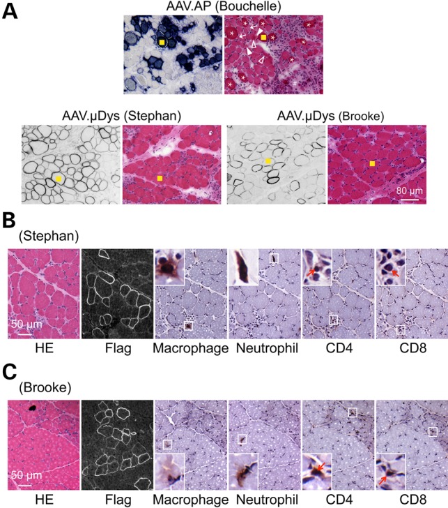
The ultimate goal of muscular dystrophy gene therapy is to treat all muscles in the body. Global gene delivery was demonstrated in dystrophic mice more than a decade ago using adeno-associated virus (AAV). However, translation to affected large mammals has been challenging. The only reported attempt was performed in newborn Duchenne muscular dystrophy (DMD) dogs. Unfortunately, AAV injection resulted in growth delay, muscle atrophy and contracture. Here we report safe and bodywide AAV delivery in juvenile DMD dogs. Three ∼2-m-old affected dogs received intravenous injection of a tyrosine-engineered AAV-9 reporter or micro-dystrophin (μDys) vector at the doses of 1.92-6.24 × 10(14) viral genome particles/kg under transient or sustained immune suppression. DMD dogs tolerated injection well and their growth was not altered. Hematology and blood biochemistry were unremarkable. No adverse reactions were observed. Widespread muscle transduction was seen in skeletal muscle, the diaphragm and heart for at least 4 months (the end of the study). Nominal expression was detected in internal organs. Improvement in muscle histology was observed in μDys-treated dogs. In summary, systemic AAV gene transfer is safe and efficient in young adult dystrophic large mammals. This may translate to bodywide gene therapy in pediatric patients in the future.


Figures

Figure 1.
Figure 1.
Experimental protocol, growth curve, blood biochemistry and biopsy results. (A) Schematic overview of the study. Arrow, AAV injection; open arrowhead, biopsy; filled arrowhead, necropsy; filled box, the dog is under immune suppression. (B) The growth curve of individual dogs. The colony average is shown in gray (N ≥ 70). (C) Selected blood biochemistry results. Complete data can be found in Supplementary Material, Table S1. Dotted gray lines, the maximal and minimal values for age-matched untreated DMD dogs in our colony (N = 31). Solid gray line, the average value of age-matched untreated DMD dogs in our colony (N = 31). (D) Representative AP histochemical staining photomicrographs and dystrophin immunofluorescence staining photomicrographs from the CT and BF muscles obtained at biopsy. (E) Vector genome copy number quantification from biopsied muscle samples.
Figure 2.
Figure 2.
A single intravenous injection of an AP reporter AAV vector leads to global muscle transduction in a juvenile dystrophic dog. (A) Representative full-view AP staining photographs of the indicated muscle. (B) Representative AP histochemical staining photomicrographs of the heart and skeletal muscles. (C) AP activity. (D) AAV vector genome quantification. Abbreviations for the heart: LA, left atrium; LVa, left ventricle anterior; LVp, left ventricle posterior; LVx, left ventricle apex; Pap, papillary muscle; RA, right atrium; RV, right ventricle; Sep, interventricular septum. Abbreviations for the diaphragm: Dia-c, diaphragm costal part; Dia-l, diaphragm lumbar part; Dia-s, diaphragm sternal part. Abbreviations for skeletal muscles: BB, biceps brachii; BF, biceps femoris; Bra, brachialis; CT, cranial tibialis; Del, deltoideus; ECU, extensor carpi ulnaris; FCR, flexor carpi radialis; FCU, flexor carpi ulnaris; FD, flexor digitorum; Gas, gastrocnemius; Gra, gracilis; Ics, intercostalis; Lat, latissimus; Pec, pectoralis; Pro, pronator; Ssp, supraspinatus; TB, triceps brachii; Ter, Teres; Ton, tongue; VL, vastus lateralis; VM, vastus medialis.
Figure 3.
Figure 3.
Systemic Y731F AAV-9 injection results in minimal transgene expression in internal organs despite robust expression in peripheral nerve, spinal cord and microvasculature. (A) Representative HE and AP histochemical staining photomicrographs of the liver, testis and kidney. (B) Representative HE and AP histochemical staining photomicrographs of the pancreas and lung. Asterisk, smooth muscle. (C) Representative AP histochemical staining photomicrographs of muscle revealing strong staining in the microvasculuture (red arrows). All arrow bars stand for 100 μm. (D) Representative AP histochemical staining photomicrographs of the sciatic never. Left panel, full-view image; right panel, high magnification image. (E) Representative HE and AP histochemical staining photomicrographs of a small never branch (arrow) inside muscle, spinal cord (gray matter and white matter), hippocampus, cerebrum and cerebellum. (F) AP activity and AAV vector genome quantification in the indicated tissues.
Figure 4.
Figure 4.
A single intravenous injection results in bodywide muscle expression of a flag-tagged μDys gene in young adult dystrophic dogs. (A) Representative flag immunostaining of skeletal muscles from dog Stephan. (B) Representative flag immunostaining of skeletal muscles from dog Brooke. (C) Representative flag immunostaining of the heart from dog Stephan. (D) Representative flag immunostaining of the heart from dog Brooke. (E) Percentage of μDys-positive myofibers. (F) Representative dystrophin western blot from dog Stephan. Positive and negative western blot controls are ECU muscles with and without μDys AAV injection, respectively. (G) Representative dystrophin western blot from dog Brooke. Positive and negative western blot controls are ECU muscles with and without μDys AAV injection, respectively. (H) AAV vector genome quantification. Abbreviations for skeletal muscles: Abd, abdominus; BB, biceps brachii; BF, biceps femoris; Bra, brachialis; CT, cranial tibialis; Del, deltoideus; Dia, diaphragm; ECU, extensor carpi ulnaris; EDL, extensor digitorum longus; EOM, extraocular muscle; FCR, flexor carpi radialis; FCU, flexor carpi ulnaris; fl-Dys, full-length dystrophin; Gas, gastrocnemius; Gra, gracilis; Ics, intercostalis; Pec, pectoralis; Pro, pronator; Sem, semitendinosus; Ssp, supraspinatus; TB, triceps brachii; Ter, Teres; Ton, tongue; VL, vastus lateralis; VM, vastus medialis. Abbreviations for the heart: LA, left atrium; LV, left ventricle; LVa, left ventricle anterior; LVp, left ventricle posterior; LVx, left ventricle apex; Pap, papillary muscle; Papa, anterior papillary muscle; Papp, posterior papillary muscle; RA, right atrium; RV, right ventricle; Rvca, caudal part of the right ventricle; RVcr, cranial part of the right ventricle; Sep, interventricular septum.
Figure 5.
Figure 5.
μDys expression ameliorates muscle pathology in young adult dystrophic dogs. (A) Representative muscle serial sections stained for transgene expression (AP staining for AAV.AP injected dog Bouchelle and flag immunostaining for AAV.μDys injected dog Stephan and Brooke) and general morphology (HE staining). The yellow square marks the same myofiber in serial sections. Asterisk, heavily stained hylinated/hypercontracted myofiber; arrow, angular myofiber; solid arrowhead, necrotic myofiber; open arrowhead, degenerative myofiber; pound sign, fibrotic tissue deposition. An enlarged view of the HE stained photomicrography of the AAV.AP infected dog is shown in Supplementary Material, Figure S3. (B) Representative HE staining, Flag immunostaining and immune cell immunohistochemical staining on muscle serial sections from AAV μDys injected dog Stephan. Insets are the enlarged view of the boxed areas. Arrow, CD4+ or CD8+ T cell. (D) Representative HE staining, Flag immunostaining and immune cell immunohistochemical staining on muscle serial sections from AAV μDys injected dog Brooke. Insets are the enlarged view of the boxed areas. Arrow, CD4+ or CD8+ T cell.

References

    1. Mingozzi F., High K.A. (2011) Therapeutic in vivo gene transfer for genetic disease using AAV: progress and challenges. Nat. Rev. Genet., 12, 341–355. - PubMed
    1. Lewis R. (2014) Gene therapy's second act. Sci. Am., 310, 52–57. - PubMed
    1. Kunkel L.M. (2005) 2004 William Allan award address. Cloning of the DMD gene. Am. J. Hum. Genet., 76, 205–214. - PMC - PubMed
    1. Wilson J.M. (2009) Lessons learned from the gene therapy trial for ornithine transcarbamylase deficiency. Mol. Genet. Metab., 96, 151–157. - PubMed
    1. Kornegay J.N., Li J., Bogan J.R., Bogan D.J., Chen C., Zheng H., Wang B., Qiao C., Howard J.F. Jr., Xiao X. (2010) Widespread muscle expression of an AAV9 human mini-dystrophin vector after intravenous injection in neonatal dystrophin-deficient dogs. Mol. Ther., 18, 1501–1508. - PMC - PubMed

Publication types